
Page 48 - Computational Retinal Image Analysis
P. 48
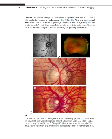
38     CHAPTER 3  The physics, instruments and modalities of retinal imaging


                         (600–800 nm) the low absorption coefficient of oxygenated blood means that arteri-
                         oles exhibit low contrast in fundus images (Fig. 5, Fig. 12c(A)) and in non-confocal
                         SLOs (Fig. 12c), but contrast is quite high in confocal-SLO images (Fig. 12a and
                         c(A)). It should be noted that a small pinhole also restricts the axial range (depth of
                         field) for detection of light to provide sectioning and profiling of the retina.


                              (a)                        (b)


                              (c)
                         FIG. 12
                         (a) Infrared (820 nm) reflectance image recorded with Heidelberg Spectralis [33]. (b) Widefield
                         two-wavelength ultra-widefield image recorded with ultrawidefield Optos SLO. (c) (A) Fundus-
                         camera photograph and (B) color SLO image [43]. Notable features are the ‘silver wiring’ in
                         image (a) and the different color and renditions and spatial resolutions of the two images in (c).