
Page 53 - Computational Retinal Image Analysis
P. 53
3 Ophthalmic instruments 43
now available from three channels, an enhanced diagnostic tool was created facilitat-
ing internal correction of movement artifacts within en-face and B-scan OCT images
using information provided by the SLO channel [50]. The efficiency of a multimodal,
multi-depth imaging tools was successfully demonstrated in various clinical studies,
an example showing simultaneously OCT, SLO and ICG images of a patient with a
well-defined classic choroidal neovascular membrane in the post-injection phase is
demonstrated in Fig. 18 [50a].
3.8.2 Spectral domain optical coherence tomography
The key value of spectral (Fourier) domain OCT is its ability to encode spatial or
temporal data into the spectrum at the interferometer output. Currently, there are
two modalities on transducing this information from the optical domain into electri-
cal: camera based (CB)-OCT where, as in TD-OCT, a broadband optical source is
employed together with a spectrometer and swept source (SS)-OCT, where a tune-
able (swept) laser is used and signal is delivered by a photo-detector. When produc-
ing B-scans, SD-OCT is clearly superior to conventional TD-OCT, in terms of both
sensitivity and acquisition speed. However, en-face imaging is still of high interest
in ophthalmology as it can often offer enhanced visualization and additional infor-
mation on sample microstructures, due to additional information conveyed that en-
hances the physician's understanding of the underlying pathology [51]. Also, en-face
imaging can be used for absolute position registration of the individual B-scan image
[52]. When implemented with TD-OCT systems, en-face OCT imaging does not al-
low reasonably high-speed data acquisition to mitigate motion artifacts [53] as they
require a quite fast phase modulation procedure in the reference arm which can be
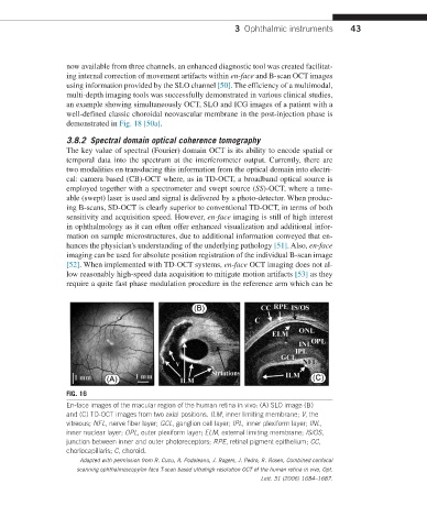
FIG. 16
En-face images of the macular region of the human retina in vivo: (A) SLO image (B)
and (C) TD-OCT images from two axial positions. ILM, inner limiting membrane; V, the
vitreous; NFL, nerve fiber layer; GCL, ganglion cell layer; IPL, inner plexiform layer; INL,
inner nuclear layer; OPL, outer plexiform layer; ELM, external limiting membrane; IS/OS,
junction between inner and outer photoreceptors; RPE, retinal pigment epithelium; CC,
choriocapillaris; C, choroid.
Adapted with permission from R. Cucu, A. Podoleanu, J. Rogers, J. Pedro, R. Rosen, Combined confocal
scanning ophthalmoscopy/en face T-scan based ultrahigh resolution OCT of the human retina in vivo, Opt.
Lett. 31 (2006) 1684–1687.