
Page 52 - Computational Retinal Image Analysis
P. 52
42 CHAPTER 3 The physics, instruments and modalities of retinal imaging
image, a B-scan image constructed of for example 500 A-scans is produced in 5 s,
and a 3D volume in which the size of the C-scans is 500 × 500 pixels in 2500 s, mak-
ing the axial TD-OCT unusable for in vivo imaging of the human eye. Another way
of producing images in TD-OCT is, for a given position of TS, to produce a lateral
reflectivity profile by transversally scanning the beam over the sample. These pro-
files (T-scans) can be used to generate cross-sections by producing them for various
values of the OPD (T-scan based B-scans). If, for a given axial position z i , T-scans
for various y j positions are assembled, a C-scan or en-face image is produced. The
advantage of this procedure is that an en-face image, at a constant depth can be pro-
duced directly, much faster, as there is no need to build the whole 3D volume. This
technique has the advantage that the quality of the image is not affected by repeated
movements of the translation stage. Thus, good quality en-face TD-OCT images can
be produced in real-time in as fast as 0.5 Hz.
Another advantage of the en-face TD-OCT is that it can be complemented by
other imaging techniques. Thus, multimodality imaging instruments were reported.
In Fig. 16A, examples of Scanning Laser Ophthalmoscopy (SLO) of the human ret-
ina and two en-face OCT images, collected from two axial positions are presented
[46]. There is pixel to pixel correspondence between the three images. The SLO
image has a poorer axial resolution, but it can serve for guidance purposes. Further
progress in the development of the multimodality imaging of human retina allowed
for sequential [47] and/or simultaneous [48] display of the en-face SLO and TD-
OCT images. Efforts were also made to increase the number of en-face images si-
multaneously displayed [49] as illustrated in Fig. 17.
In addition, the multichannel potential of the OCT/SLO system was demon-
strated by the addition of a third hardware channel which acquires and generates
indocyanine green (ICG) fluorescence images. Thus, three en-face images, SLO,
OCT and ICG fluorescence images were simultaneously presented and the synergy
between the simultaneously provided perspectives demonstrated. With information
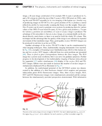
FIG. 15
Procedure for producing a B-scan image in TD-OCT. For each lateral position x p , p = 1..P,
the signal I(x p ,z q ) (q = 1..Q) is collected for each position z q of the translation stage TS.