
Page 46 - Computational Retinal Image Analysis
P. 46
36 CHAPTER 3 The physics, instruments and modalities of retinal imaging
distance, and (generally) increase the field of view, although the latter depends on the
diameter of the lens. Because the observed image is inverted, use of the device for
navigation is not intuitive and requires some training (Fig. 11).
3.5 The scanning laser ophthalmoscopes
The Scanning Laser Ophthalmoscope (SLO) is a fundamentally different approach
to recording retinal images involving the scanning of a laser spot across the retina.
An image is formed by the detection of light that is backscattered through the pupil,
through the SLO optics to a detector. This provides a measurement of the fundus
reflectance at the particular point of the illumination spot. In contrast to the snapshot
image acquisition by the fundus camera, raster scanning of the laser across the retina
to record a 2D image can take as long as a second, which can lead to image artifacts
if the eye moves during recording.
Interestingly, the SLO illumination concept reverses the Gullstrand principle
of illumination: a narrow laser beam (typically <1 mm) is transmitted through the
center of the pupil and the rest of the pupil is used to collect backscattered light
[39, 40]. This inversion of the illumination-imaging light paths enables a larger area
of the pupil to be dedicated to the collection of light, increasing optical efficiency.
Fundamentally, it is possible to reduce the illumination area only because of the
higher intensity of lasers compared to thermal light sources. A penalty for the use of
the smaller beam is that it yields a smaller angular and spatial resolution: according
to Eq. (1), this yields a spatial resolution of about 10 μm at the retina, which is insuf-
ficient to resolve individual blood cells and the smaller capillaries.
As can be seen from a comparison of the images in Fig. 12, images recorded
with a SLO can look strikingly different from those recorded with a broadband light
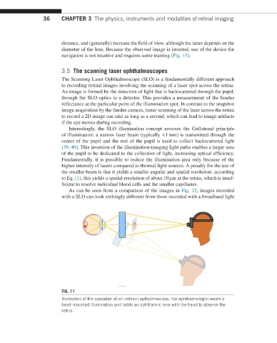
FIG. 11
Illustration of the operation of an indirect opthalmoscope, the ophthalmologist wears a
head-mounted illumination and holds an ophthalmic lens with the hand to observe the
retina.