
Page 42 - Computational Retinal Image Analysis
P. 42
32 CHAPTER 3 The physics, instruments and modalities of retinal imaging
the green (532 nm) and red (633 nm) wavelengths it is notable that the hybrid multi-
mode images, which employ an agile combination of confocal and direct imaging,
yield images of the vasculature with increased contrast. This is particularly true for
the arterioles, which are not visible in the direct-mode images at the red wavelengths.
Also notable is that the choriocapillaris is visible in the red images, due to the higher
transparency, but not in the green images (Fig. 8).
2.6 The use of eye phantoms to simulate retinal imaging
Eye phantoms are commonly used to perform experimental, simulation tests and vali-
dation. To model the optics of the eye as an imaging system, several simulation models
have been proposed, called schematic eye models [8, 9]. They take into account the
geometric shapes and index of refraction of each ocular component: the cornea, lens,
vitreous humor and aqueous humor, with varying degree of rigor. These models enable
optical simulation based on ray-tracing, and thus they can be inserted in the design of
ophthalmic instruments to model and optimize their performance, and to account for
the effects of the eye such as aberrations, optical scatter by the ocular media and diffrac-
tion. These models range from simple approximations, based on simpler geometrical
shapes and models of refractive index, to more complete descriptions that may include
accurate chromatic dispersion, aspheric surfaces and graded-index (GRIN) models, bi-
refringence, and other properties [7], and may describe better the optical performance
on a wider range of angles (e.g., for a wide field-of-view). For experimental assessment
and validation, phantom eyes can be built to mimic the eye such as are shown in Fig. 9.
These range from simple models based on a single lens and a flat target resembling the
retina, to more complex and accurate models based on a lens pair mimicking the cornea
and crystalline lens, enclosed in a case filled with water and a curved and layered target
to mimic volumetric scattering at the retina [35, 36].
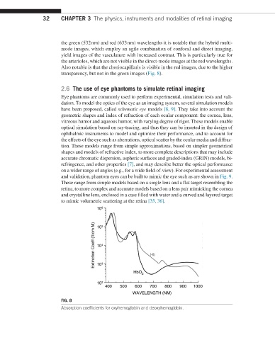
FIG. 8
Absorption coefficients for oxyhemoglobin and deoxyhemoglobin.