
Page 99 - Computational Retinal Image Analysis
P. 99
Summary 91
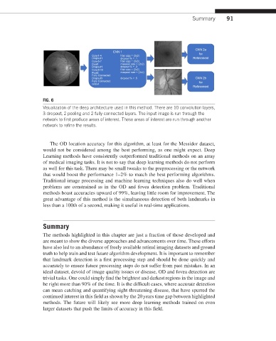
FIG. 6
Visualization of the deep architecture used in this method. There are 10 convolution layers,
3 dropout, 2 pooling and 2 fully connected layers. The input image is run through the
network to first produce areas of interest. These areas of interest are run through another
network to refine the results.
The OD location accuracy for this algorithm, at least for the Messidor dataset,
would not be considered among the best performing, as one might expect. Deep
Learning methods have consistently outperformed traditional methods on an array
of medical imaging tasks. It is not to say that deep learning methods do not perform
as well for this task. There may be small tweaks to the preprocessing or the network
that would boost the performance 1–2% to match the best performing algorithms.
Traditional image processing and machine learning techniques also do well when
problems are constrained as in the OD and fovea detection problem. Traditional
methods boast accuracies upward of 99%, leaving little room for improvement. The
great advantage of this method is the simultaneous detection of both landmarks in
less than a 100th of a second, making it useful in real-time applications.
Summary
The methods highlighted in this chapter are just a fraction of those developed and
are meant to show the diverse approaches and advancements over time. These efforts
have also led to an abundance of freely available retinal imaging datasets and ground
truth to help train and test future algorithm development. It is important to remember
that landmark detection is a first processing step and should be done quickly and
accurately to ensure future processing steps do not suffer from past mistakes. In an
ideal dataset, devoid of image quality issues or disease, OD and fovea detection are
trivial tasks. One could simply find the brightest and darkest regions in the image and
be right more than 90% of the time. It is the difficult cases, where accurate detection
can mean catching and quantifying sight-threatening disease, that have spurred the
continued interest in this field as shown by the 20 years time gap between highlighted
methods. The future will likely see more deep learning methods trained on even
larger datasets that push the limits of accuracy in this field.