
Page 417 -
P. 417
Section 12.3 Registering Deformable Objects 385
Image 1 Rigid Affine Deformation
Image 2
Difference
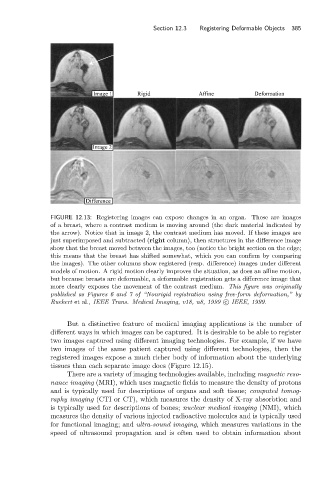
FIGURE 12.13: Registering images can expose changes in an organ. These are images
of a breast, where a contrast medium is moving around (the dark material indicated by
the arrow). Notice that in image 2, the contrast medium has moved. If these images are
just superimposed and subtracted (right column), then structures in the difference image
show that the breast moved between the images, too (notice the bright section on the edge;
this means that the breast has shifted somewhat, which you can confirm by comparing
the images). The other columns show registered (resp. difference) images under different
models of motion. A rigid motion clearly improves the situation, as does an affine motion,
but because breasts are deformable, a deformable registration gets a difference image that
more clearly exposes the movement of the contrast medium. This figure was originally
published as Figures 6 and 7 of “Nonrigid registration using free-form deformation,” by
Ruekert et al., IEEE Trans. Medical Imaging, v18, n8, 1999 c IEEE, 1999.
But a distinctive feature of medical imaging applications is the number of
different ways in which images can be captured. It is desirable to be able to register
two images captured using different imaging technologies. For example, if we have
two images of the same patient captured using different technologies, then the
registered images expose a much richer body of information about the underlying
tissues than each separate image does (Figure 12.15).
There are a variety of imaging technologies available, including magnetic reso-
nance imaging (MRI), which uses magnetic fields to measure the density of protons
and is typically used for descriptions of organs and soft tissue; computed tomog-
raphy imaging (CTI or CT), which measures the density of X-ray absorbtion and
is typically used for descriptions of bones; nuclear medical imaging (NMI), which
measures the density of various injected radioactive molecules and is typically used
for functional imaging; and ultra-sound imaging, which measures variations in the
speed of ultrasound propagation and is often used to obtain information about