
Page 415 -
P. 415
Section 12.3 Registering Deformable Objects 383
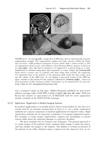
FIGURE 12.11: On the top left, a single slice of MRI data with an automatically acquired
segmentation overlaid. The segmentation outlines the brain, vacuoles within the brain,
and the tumor. MRI produces a sequence of slices, which yield a volume model; a view
of a segmented volume model, with different colors showing different regions, is shown at
the top right. Once this data is obtained, it is registered to a patient lying on a table.
Registration is obtained using depth data measured by a laser ranger; the bottom-left
figure shows a camera view of a patient with laser ranger data overlaid. By registering
the segmented data to the patient on the operating table using this laser ranger data
and the surface of the MRI data, we can display a processed version of the MRI im-
agery overlaid on the patient for the surgeon’s information (bottom right). Figures by
kind permission of Eric Grimson; further information can be obtained from his web site,
http://www.ai.mit.edu/people/welg/welg.html.
and a beginner’s guide on this page. Mikkel Stegmann publishes an open-source
software package called AAM-API at http://www2.imm.dtu.dk/ ~ aam/. Dirk-Jan
Kroon has released an open-source set of MATLAB tools for active appearance
models, available at the MATLAB file exchange.
12.3.3 Application: Registration in Medical Imaging Systems
In medical applications, it is usually known what is being looked at, but there is a
crucial need for an accurate measurement of where it is. As a result, registration
methods are a major component of medical applications of computer vision. Rigid
registration methods are an important component of computer-supported surgery.
For example, in brain surgery applications, surgeons are attempting to remove
tumors while doing the minimum damage to a patient’s faculties.
We show examples due to Grimson and colleagues. The general approach is
to obtain images of the patient’s brain, segment these images to show the tumor,
and then display the images to the surgeon. The display is overlaid on pictures of
the patient on the table, obtained using a camera near the surgeon’s view, to cue