
Page 419 -
P. 419
Section 12.3 Registering Deformable Objects 387
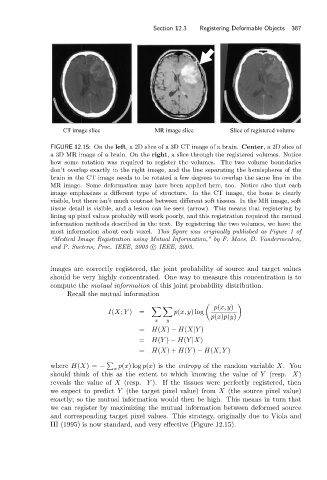
CT image slice MR image slice Slice of registered volume
FIGURE 12.15: On the left, a 2D slice of a 3D CT image of a brain. Center,a2Dslice of
a 3D MR image of a brain. On the right, a slice through the registered volumes. Notice
how some rotation was required to register the volumes. The two volume boundaries
don’t overlap exactly in the right image, and the line separating the hemispheres of the
brain in the CT image needs to be rotated a few degrees to overlap the same line in the
MR image. Some deformation may have been applied here, too. Notice also that each
image emphasizes a different type of structure. In the CT image, the bone is clearly
visible, but there isn’t much contrast between different soft tissues. In the MR image, soft
tissue detail is visible, and a lesion can be seen (arrow). This means that registering by
lining up pixel values probably will work poorly, and this registration required the mutual
information methods described in the text. By registering the two volumes, we have the
most information about each voxel. This figure was originally published as Figure 1 of
“Medical Image Registration using Mutual Information,” by F. Maes, D. Vandermeulen,
and P. Suetens, Proc. IEEE, 2003 c IEEE, 2003.
images are correctly registered, the joint probability of source and target values
should be very highly concentrated. One way to measure this concentration is to
compute the mutual information of this joint probability distribution.
Recall the mutual information
p(x, y)
I(X; Y ) = p(x, y)log
p(x)p(y)
x y
= H(X) − H(X|Y )
= H(Y ) − H(Y |X)
= H(X)+ H(Y ) − H(X, Y )
where H(X)= − x p(x)log p(x)is the entropy of the random variable X.You
should think of this as the extent to which knowing the value of Y (resp. X)
reveals the value of X (resp. Y ). If the tissues were perfectly registered, then
we expect to predict Y (the target pixel value) from X (the source pixel value)
exactly; so the mutual information would then be high. This means in turn that
we can register by maximizing the mutual information between deformed source
and corresponding target pixel values. This strategy, originally due to Viola and
III (1995) is now standard, and very effective (Figure 12.15).