
Page 136 - Academic Press Encyclopedia of Physical Science and Technology 3rd BioTechnology
P. 136
P1: LEF/GUR Final Pages P2: GTV
Encyclopedia of Physical Science and Technology EN007O-865 July 6, 2001 17:0
Image-Guided Surgery 585
apy methods. The availability of patient registered, con-
tinuously updated “fused” multimodal information in an
intraoperative setting increases safety and may result in
better outcome by reducing the invasiveness of the proce-
dures, decreasing complications, and increasing the effec-
tiveness of surgery. Image-based information can be uti-
lized accurately to target and cut out diseased tissues and
at the same time avoid critical structures. During surgery
most of the structures and the related functions are unseen
by the surgeon but can be displayed interactively.
Intraoperative shifts and deformations are unavoidable
and mostly unpredictable. These displacements are the
results of mechanical factors, physiologic motions, and
pathophysiologic processes like edema or hemorrhage.
The unwanted movement of tissues and the reduction or
swellingoftissuevolumesbytheadvancingsurgerycanbe
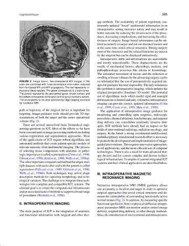
FIGURE 2 Image fusion. Two-dimensional MR images of the so substantial that the use of preoperatively acquired im-
brain are combined with three-dimensional information obtained ages for guidance become impossible. The only solution to
from functional MR and MR angiograms. The red represents in-
this problem is intraoperative imaging, which updates the
tracranial blood vessels. The green corresponds to a brain tumor.
original preoperative (baseline) 3D model. The potential
The purple represents the pre-central gyrus (motor cortex) and
the yellow corresponds to the post-central gyrus (sensory cortex). use of algorithmic tools which model rigid and nonrigid
The arrow points to the area activated by finger tapping recorded deformations is limited and only volumetric intraoperative
by functional MRI. imaging can provide correct, updated information (Cotin
et al., 1999; Cover et al., 1993; Hata et al., 1998).
path or trajectory of the surgical device is important for The application of intraoperative image guidance for
targeting. Image-guidance tools should provide 3D rep- monitoring and controlling open surgeries, endoscopic
resentations of both the target and the entire operational procedures,thermalablations,brachytherapy,andtargeted
volume (Fig. 2). drug delivery can consolidate minimally invasive ther-
There are several unresolved basic biomedical engi- apies. IGT methods have already had an impact on the
neering questions in IGT. Most of the efforts so far have fields of interventional radiology, radiation oncology, and
been concentrated on image processing methods including surgery. In the future a strong coordinated multifocused,
various registration and segmentation approaches. Most multidisciplinary translational research effort is necessary
of the applications of IGT require robust algorithms and topromotethedevelopmentandimplementationofimage-
automated methods that create patient-specific models of guidedinterventions.Thisrequiresinnovativeapproaches,
relevant anatomy from multimodal imaging. The process novel applications, and the more efficient use of computer
of selecting tissue components with anatomic or patho- technologies. There is also a need for more advanced ther-
logicimportanceiscalledsegmentation(Clineetal.,1990; apy devices and for a more complex and diverse techno-
Gibson et al., 1998; Held et al., 1996; Wells et al., 1996a). logical infrastructure. Examples of current integrated IGT
The other important computerized method that aligns mul- systems and their clinical application are described below.
tiple datasets with each other and with the patient is called
registration (Pelizzari et al., 1989; Grimson et al., 1996;
Wells et al., 1996b). Both techniques may utilize shape III. INTRAOPERATIVE MAGNETIC
description methods for capturing morphology and its bi- RESONANCE IMAGING
ological variation. The challenge is to integrate these tech-
nologies into complete and compatible IGT systems. The
Interactive intraoperative MRI (IMRI) guidance allows
ultimate goal is to create the computational infrastructure one accurately to localize and target in order to optimize
andanassociatedsuiteofmethodstosupportabroadrange surgical approaches that avoid critical structures and de-
of procedures (Warfield et al., 1998). crease the vulnerability of surrounding functionally active
normal tissues (Fig. 2). In addition, by measuring specific
II. INTRAOPERATIVE IMAGING functional(perfusion,flow)orphysical(diffusion,temper-
ature) parameters MRI can monitor and/or control energy
The main purpose of IGT is the integration of anatomic delivery, targeted drug delivery, or other therapy methods.
and functional information with surgical and other ther- Since the introduction of interventional and intraoperative