
Page 139 - Academic Press Encyclopedia of Physical Science and Technology 3rd BioTechnology
P. 139
P1: LEF/GUR Final Pages P2: GTV
Encyclopedia of Physical Science and Technology EN007O-865 July 6, 2001 17:0
588 Image-Guided Surgery
be developed to utilize dynamic imaging methods, such as
MR fluoroscopy, keyhole imaging, and adaptive imaging.
Since its initial introduction IMRI has matured from a
research tool into a clinical approach that can transform
minimally invasive surgery and interventional radiology
into a more advanced stage. Currently several open-
configuration and short-bore MRI systems suitable for
some percutaneous procedures and/or open surgeries are
being marketed. In areas like neurosurgery and endo-
scopic surgery, MRI-based guidance systems may pro-
vide more effective treatment options than conventional
surgery or other image-guidance techniques such as ul-
trasound and X-ray computed tomography. Intraoperative
MRI may result in improved patient care, reduced inva-
siveness, and a safer surgical or interventional procedure.
MRI-based image guidance may induce the development
and implementation of new surgical approaches. This is
a challenging new technology which can lead to signifi-
cant changes in surgical procedures and other treatment
methods.
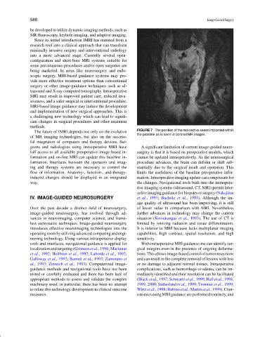
FIGURE 7 The position of the radioactive seeds implanted within
The future of IMRI depends not only on the evolution
the prostate as is seen in coronal MR images.
of MR imaging technologies, but also on the success-
ful integration of computers and therapy devices. Sur-
geons and radiologists using intraoperative MRI have A significant limitation of current image-guided neuro-
full access to all available preoperative image based in- surgery is that it is based on preoperative models, which
formation and on-line MRI can update this baseline in- cannot be updated intraoperatively. As the neurosurgical
formation. Interfaces between the operators and imag- procedure advances, the brain can deform or shift sub-
ing and therapy systems are necessary to control the stantially due to the surgical insult and operation. This
flow of information. Anatomy-, function-, and therapy- limits the usefulness of the baseline preoperative infor-
induced changes should be displayed in an integrated mation. Intraoperative imaging update can compensate for
way. the changes. Navigational tools built into the intraopera-
tive imaging systems (ultrasound, CT, MRI) permit inter-
active imaging guidance for biopsies or surgery (Nakajima
IV. IMAGE-GUIDED NEUROSURGERY et al., 1997; Bucholz et al., 1993). Although the im-
age quality of ultrasound has been improving, it is still
Over the past decade a distinct field of neurosurgery, of lesser value in comparison with MRI. Nevertheless
image-guided neurosurgery, has evolved through ad- further advances in technology may change the current
vances in neuroimaging, computer science, and frame- situation (Koivukangas et al., 1993). The use of CT is
less stereotactic techniques. Image-guided neurosurgery limited by ionizing radiation and tissue differentiation.
introduces effective neuroimaging technologies into the It is inferior to MRI because lacks multiplanar imaging
operating room by utilizing advanced computing and engi- capabilities, high contrast, spatial resolution, and high
neering technology. Using various intraoperative display sensitivity.
tools and interfaces, navigational guidance is applied for With intraoperative MRI guidance one can identify sur-
localization and targeting (Grimson et al., 1996; Maciunas gical margins even in the presence of ongoing deforma-
et al., 1992; Heilbrun et al., 1992; Laborde et al., 1992; tions. This allows image-based control of tumor resections
Galloway et al., 1992; Barnett et al., 1993; Zamorano et and can result in the complete removal of lesions with less
al., 1993; Zinreich et al., 1993). Computerized image- or no damage to adjacent normal tissues. Intraoperative
guidance methods and navigational tools have not been complications, such as hemorrhage or edema, can be im-
tested or carefully evaluated and there has been lack of mediately identified and their resolution can be facilitated
appropriate methods to assess and validate the complex (Black et al., 1997; Schwartz et al., 1999; Hall et al., 1998,
machinery used; in particular, there has been no attempt 1999, 2000; Sutherland et al., 1999; Tronnier et al., 1999;
to relate this technology development to clinical outcome Wirtz et al., 1998; Rubino et al., Martin et al., 1999). Cran-
measures. iotomies using MRI guidance are performed routinely, and