
Page 142 - Academic Press Encyclopedia of Physical Science and Technology 3rd BioTechnology
P. 142
P1: LEF/GUR Final Pages P2: GTV
Encyclopedia of Physical Science and Technology EN007O-865 July 6, 2001 17:0
Image-Guided Surgery 591
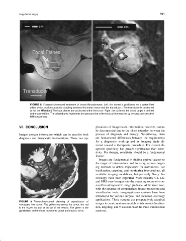
FIGURE 8 Focused ultrasound treatment of breast fibroadenoma. Left: the breast is positioned on a water-filled
pillow which provides acoustic coupling between the breast tissue and the transducer. (The transducer is positioned
within the MR table.) The focal planes are positioned within the tumor. Right: the outline of the tumor target is defined
by the dashed line. The colored area represents temperature rise at the focal point measured by temperature-sensitive
MRI sequences.
VII. CONCLUSION plications of image-based information, however, cannot
be disconnected due to the close interplay between the
Images contain information which can be used for both process of diagnosis and therapy. Nevertheless, there
diagnosis and therapeutic interventions. These two ap- are fundamental differences between the requirements
for a diagnostic work-up and an imaging study di-
rected toward a therapeutic procedure. For correct di-
agnosis specificity has greater significance than sensi-
tivity. For therapy, sensitivity should be a fundamental
feature.
Images are fundamental in finding optimal access to
the target of interventions and in using various target-
ing methods to define trajectories for instruments. For
localization, targeting, and monitoring interventions, all
available imaging modalities, but primarily X-ray flu-
oroscopy, have been exploited. More recently CT, US,
and MRI were brought into the operating room environ-
ment for intraoperative image guidance. At the same time,
with the advance of computerized image processing and
visualization tools, image-guidance systems have been
introduced for various surgical and radiation oncology
applications. These systems use preoperatively acquired
FIGURE 9 Three-dimensional planning of cryoablation of
metastatic liver tumor. The yellow represents the tumor, the red images to create anatomic models which provide localiza-
is the frozen ice ball at the tip of the needle. The green is the tion, targeting, and visualization of the three-dimensional
gallbladder and the blue represents portal and hepatic veins. anatomy.