
Page 137 - Academic Press Encyclopedia of Physical Science and Technology 3rd BioTechnology
P. 137
P1: LEF/GUR Final Pages P2: GTV
Encyclopedia of Physical Science and Technology EN007O-865 July 6, 2001 17:0
586 Image-Guided Surgery
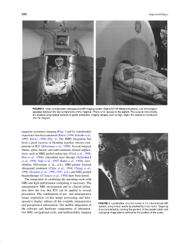
FIGURE 3 Open-configuration intraoperative MR imaging system (Signa SP, GE Medical Systems). Left: the surgeon
operates between the two components of the magnets. There is full access to the patient. The surgical instruments
are tracked using optical sensors to guide interactive imaging (display seen at top). Right: the patient is introduced
into the magnet.
magnetic resonance imaging (Figs. 3 and 4), considerable
experience has been amassed (Jolesz, 1994; Schenk et al.,
1995; Jolesz, 1998) (Fig. 4). The IMRI integration has
been a good exercise in blending together various com-
ponents of IGT (Silverman et al., 1998). Several surgical
(brain, spine, breast) and interventional clinical applica-
tions, such as MRI-guided endoscopy (Fried et al., 1998;
Hsu et al., 1998), interstitial laser therapy (Kettenbach
et al., 1998; Vogl et al., 1997; Kahn et al., 1998), cryo-
ablation (Silverman et al., n.d.), MRI-guided focused
ultrasound treatment (Cline et al., 1994; Chung et al.,
1996; Hynynen et al., 1996, 1997, n.d.), and MRI-guided
brachytherapy (D’Amico et al., 1998) have been tested.
The integration of combining the operating room with
MRI and high-performance computing is necessary. The
intraoperative MRI environment and its clinical utiliza-
tion show the way that IGT can be applied to several
procedures. The combination of pre- and intraoperative
image acquisition, on-line image processing, and intra-
operative display utilizes all the available intraoperative
FIGURE 4 Localization of a liver tumor in the interventional MR
and preoperative information. The skillful integration of
system, using virtual needle tip (marked by cross-hairs). Targeting
the software and hardware components of intraopera- is accomplished by tracking the position of the needle holder and
tive MRI, navigational tools, and multimodality imaging displaying image planes defined by the position of the probe.