
Page 191 - Academic Press Encyclopedia of Physical Science and Technology 3rd BioTechnology
P. 191
P1: FMX Final Pages
Encyclopedia of Physical Science and Technology EN009J-69 July 19, 2001 22:50
Microanalytical Assays 691
mobilized on the interior surface membrane. This ensures
retention of both the Dextran and Con A in the sensor. In
the absence of glucose in the external ‘ medium, most of
the FITC Dextran will be occupying sites on the Con A out
of the view of the light that comes out of the distal end of
the optical fiber, and thus there is very little of fluorescence
that enters back into the optical fiber. On the other hand, if
the responsive end of the optic fiber is placed in a solution
containing sugar, glucose can diffuse through the wall of
the dialysis tubing and compete for Con A binding sites,
displacing some of the FITC Dextran that then distributes
uniformly throughout the lumen of the hollow fiber,
and the fraction in the middle of the hollow fiber is ex-
cited by the light that comes out of the optical fiber. A
portion of the emitted fluorescence from FITC Dextran is
captured by the same optical fiber producing, transmitted
to a photomultiplier that produces a signal directly related
to the glucose concentration in the external medium.
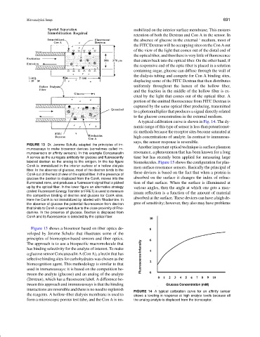
A typical calibration curve is shown in Fig. 14. The dy-
namic range of this type of sensor is less than potentiomet-
ric methods because the receptor sites become saturated at
high concentrations of analyte. In contrast to immunoas-
says, the sensor response is reversible.
FIGURE 13 Dr. Jerome Schultz adapted the principles of im-
Another important optical technique is surface plasmon
munoassays to make biosensor devices (sometimes called im-
resonance, a phenomenon that has been known for a long
munosensors or affinity sensors). In this example Concanavalin
A serves as the surrogate antibody for glucose and fluorescently time but has recently been applied for measuring large
labeled dextran as the analog to the antigen. In the top figure biomolecules. Figure 15 shows the configuration for plas-
ConA is immobilized in the interior surface of a hollow dialysis mon surface resonance sensors. Basically the principal of
fiber. In the absence of glucose, most of the dextran binds to the
ConA out of the field of view of the optical fiber. In the presence of these devices is based on the fact that when a protein is
glucose the dextran is displaced from the ConA, moves into the absorbed on the surface it changes the index of refrac-
illuminated zone, and produces a fluorescent signal that is picked tion of that surface. When the surface is illuminated at
up by the optical fiber. In the lower figure an alternative strategy various angles, then the angle at which one gets a max-
(called Fluorescent Energy Transfer or FRET) is used to measure imum reflection is a function of the amount of material
the competitive binding of dextran and glucose for ConA sites.
Here the ConA is not immobilized by labeled with Rhodamine. In absorbed at the surface. These devices can have a high de-
the absence of glucose the potential fluorescence from dextran gree of sensitivity; however, they also may have problems
that binds to ConA is quenched due to the close proximity of Rho-
damine. In the presence of glucose, Dextran is displaced from
ConA and its fluorescence is detected by the optical fiber.
Figure 13 shows a biosensor based on fiber optics de-
veloped by Jerome Schultz that illustrates some of the
principles of bioreceptor-based sensors and fiber optics.
The approach is to use a biospecific macromolecule that
has binding selectivity for the analyte of interest. To make
a glucose sensor Concanavalin A (Con A), a lectin that has
selective binding sites for carbohydrates was chosen as the
biorecognition agent. This methodology is similar to that
used in immunoassays: it is based on the competition be-
tween the analyte (glucose) and an analog of the analyte
(Dextran), which has a fluorescent label. A difference be-
tween this approach and immunoassays is that the binding
interactions are reversible and there is no need to replenish
FIGURE 14 A typical calibration curve for an affinity sensor
the reagents. A hollow-fiber dialysis membrane is used to shows a leveling-in response at high analyte levels because all
form a microscopic porous test tube, and the Con A is im- the analog analyte is displaced from the bioreceptor.