
Page 204 - Academic Press Encyclopedia of Physical Science and Technology 3rd BioTechnology
P. 204
P1: GTV/MBR P2: GSS Final Pages
Encyclopedia of Physical Science and Technology EN011L-523 August 10, 2001 11:17
324 Optical Fiber Techniques for Medical Applications
ancillary channel through which surgical instruments can
be inserted and used for endoscopic therapy or surgery or
for the removal of tissues, such as a biopsy or the removal
of tumors.
Some endoscopes are listed in alphabetical order:
Arthroscope—for the joints
Bronchoscope—for the bronchi
Colonoscope—for the colon
Colposcope—for the cervix and the vagina
Cystoscope—for the urinary bladder
Gastroscope—for the stomach, esophagus, and the
bile duct
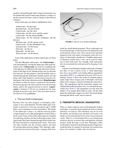
Hysteroscope—for the uterine cavity FIGURE 8 A picture of an ultrathin endoscope.
Laparoscope—for the abdominal cavity
Laryngoscope—for the larynx
Otoscope—for the ear inside the womb during pregnancy. These endoscopes can
Sinuscope—for the nose be inserted through a small incision in the abdominal wall
Thoracoscope—for the thorax. or through the uterine entry. They may provide important
diagnosis in the early stages of pregnancy, where the reso-
Some of the applications of these endoscopes are listed lution of ultrasound is insufficient. Ultrathin endoscopes,
below: of diameters smaller than 1 mm, can be used for imag-
The first fiberoptic endoscopes—the Gastroscopes— ing of other body parts. For example, rigid endoscopes
were developed for viewing the upper part of the gastroin- have been inserted into teeth and used for imaging of root
testinal tract. Colonoscopes are used for examining the canals.
colon and may be utilized for the early detection of carci- A picture of an Olympus ultrathin endoscope of diamter
noma of the large bowel. Benign polyps may be detected less than 1.0 mm is given in Fig. 8. Such endoscopes
and removed. For this purpose a special metallic snare is have been successfully used during balloon angioplasty
inserted through the instrument channel, passed over the procedures (PTCA, as explained in Section VII.E on car-
polyp, and a high-frequency current used to heat the wire diology). Figure 9 shows the results of standard angiog-
and remove the polyp (electroresection). Bronchoscopes raphy (upper three images) and of fiberoptic endoscopy
are thinner, as they are used to visualize the thin bronchi. (three lower images), carried out during the same proce-
Bronchoscopes have also been used for removal of foreign dure. The angiograms and the endoscopic images were
bodies, and for the surgical removal of tumors. Laparo- obtained before the procedure (Fig. 9A) and after the pro-
scopes of diameter 5–10 mm are inserted into the body cedure (Fig. 9B & C). The angiograms can only show the
through an incision in the navel. They have been used for shadow of an opaque fluid inside an artery. On the other
the removal of the gallbladder. hand, the endoscopic images can show the actual plaque
blocking the blood vessel, and its removal.
2. Thin and Ultrathin Endoscopes
Recently there has been progress in developing endo- V. FIBEROPTIC MEDICAL DIAGNOSTICS
scopes of very small diameter. Flexible endoscopes of di-
ameter of about 0.5–2 mm may incorporate up to 10,000 There is a need to improve some of the diagnostic medical
fibers, each of diameter of a few micrometers. The length techniques. At present, blood samples are sent to a remote
of the endoscope is about 1 m, and the resolving power laboratory for analysis. This laboratory may be far from
is high enough to see a thin polypropylene suture inside a the patient and the physician and there are bound to be
blood vessel. Some of these thin endoscopes are intended delays or even unintentional errors in the clinical chemical
for cardiology and they are then called angioscopes. Such results. There is a concentrated effort to use miniaturized
angioscopes have been inserted through blood vessels into electronic devices as sensors that could perform chemical
the heart and used for examining heart valves. They have analysis inside the body, in real time. Fiberoptics offers
also been inserted into the coronary arteries and used an alternative method for performing medical diagnostics
for viewing atherosclerotic plaque. Thin, fiberoptic endo- inside the body of a patient. In principle, this method may
scopes have been used in fetoscopy—imaging of the fetus prove to be sensitive, reliable, and cost effective.