
Page 203 - Academic Press Encyclopedia of Physical Science and Technology 3rd BioTechnology
P. 203
P1: GTV/MBR P2: GSS Final Pages
Encyclopedia of Physical Science and Technology EN011L-523 August 10, 2001 11:17
Optical Fiber Techniques for Medical Applications 323
TABLE II Typical Data on Commercially
Available Flexible Endoscopes
Length 300–2500 mm
Outer diameter 0.5–15 mm
Instrumental channel diameter 0.5–3mm
◦
Flexible up/down 180 /60 ◦
Depth of focus 5–100 mm
◦
Field of view 50 –100 ◦
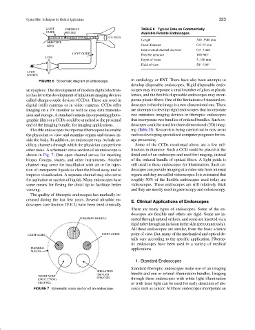
FIGURE 6 Schematic diagram of a fiberscope. in cardiology or ENT. There have also been attempts to
develop disposable endoscopes. Rigid disposable endo-
an eyepiece. The development of modern digital electron- scopes may incorporate a small number of glass or plastic
icshaslettothedevelopmentofminiatureimagingdevices lenses, and the flexible disposable endoscopes may incor-
called charge-couple devices (CCDs). These are used in porate plastic fibers. One of the limitations of standard en-
digital (still) cameras or in video cameras. CCDs offer doscopes is that the image is a two-dimensional one. There
imaging on a TV monitor as well as easy data transmis- are attempts to develop rigid endoscopes that incorporate
sion and storage. A standard camera (incorporating photo- two miniature imaging devices or fiberoptic endoscopes
graphic film) or a CCDs could be attached to the proximal that incorporate two bundles of ordered bundles. Such en-
end of the imaging bundle, for imaging applications. doscopes could be used for three-dimensional (3D) imag-
Flexible endoscopes incorporate fiberscopes that enable ing (Table II). Research is being carried out in new areas
the physician to view and examine organs and tissues in- such as developing specialized computer programs for im-
side the body. In addition, an endoscope may include an- age processing.
cillary channels through which the physician can perform Some of the CCDs mentioned above are a few mil-
other tasks. A schematic cross section of an endoscope is limeters in diameter. Such a CCD could be placed at the
shown in Fig. 7. One open channel serves for inserting distal end of an endoscope and used for imaging, instead
biopsy forceps, snares, and other instruments. Another of the ordered bundle of optical fibers. A light guide is
channel may serve for insufflation with air or for injec- still used in these endoscopes for illumination. Such en-
tion of transparent liquids to clear the blood away and to doscopes can provide imaging at a video rate from internal
improve visualization. A separate channel may also serve organs and they are called videoscopes. It is estimated that
for aspiration or suction of liquids. Many endoscopes have roughly 50% of the flexible endoscopes used today are
some means for flexing the distal tip to facilitate better videoscopes. These endoscopes are still relatively thick
viewing. and they are mostly used in gastroscopy and colonoscopy.
The quality of fiberoptic endoscopes has markedly in-
creased during the last few years. Several ultrathin en-
E. Clinical Applications of Endoscopes
doscopes (see Section IV.E.2) have been tried clinically
There are many types of endoscopes. Some of the en-
doscopes are flexible and others are rigid. Some are in-
serted through natural orifices, and some are inserted via a
rigid tube through an incision in the skin (percutaneously).
All these endoscopes are similar, from the basic science
point of view. But, many of the mechanical and optical de-
tails vary according to the specific application. Fiberop-
tic endoscopes have been used in a variety of medical
applications.
1. Standard Endoscopes
Standard fiberoptic endoscopes make use of an imaging
bundle and one or several illumination bundles. Imaging
through these endoscopes with white light illumination
or with laser light can be used for early detection of dis-
FIGURE 7 Schematic cross section of an endoscope. eases such as cancer. All these endoscopes incorporate an