
Page 14 - Academic Press Encyclopedia of Physical Science and Technology 3rd Analytical Chemistry
P. 14
P1: GJB Revised Pages
Encyclopedia of Physical Science and Technology En001f25 May 7, 2001 13:58
Analytical Chemistry 553
−1
and 750 cm , which is often called the “fingerprint” re-
gion. Such analyses can be applied to solid-, liquid-, and
gas-phase samples, and a summary of some common ab-
sorption bands useful for chemical identification purposes
is provided in Table V.
Electron spin resonance. In a strong magnetic
field, the degenerate energy levels designated by the elec-
1
tron spin quantum number ± actually differ in energy.
2
The difference in energy between these levels is described
as E,
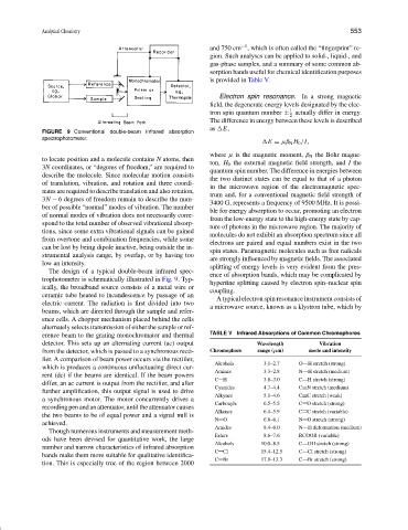
FIGURE 9 Conventional double-beam infrared absorption
spectrophotometer.
E = µβ N H 0 /I,
where µ is the magnetic moment, β N the Bohr magne-
to locate position and a molecule contains N atoms, then
ton, H 0 the external magnetic field strength, and I the
3N coordinates, or “degrees of freedom,” are required to
quantum spin number. The difference in energies between
describe the molecule. Since molecular motion consists
the two distinct states can be equal to that of a photon
of translation, vibration, and rotation and three coordi-
in the microwave region of the electromagnetic spec-
nates are required to describe translation and also rotation,
trum and, for a conventional magnetic field strength of
3N − 6 degrees of freedom remain to describe the num-
3400 G, represents a frequency of 9500 MHz. It is possi-
ber of possible “normal” modes of vibration. The number
ble for energy absorption to occur, promoting an electron
of normal modes of vibration does not necessarily corre-
from the low-energy state to the high-energy state by cap-
spond to the total number of observed vibrational absorp-
ture of photons in the microwave region. The majority of
tions, since some extra vibrational signals can be gained
molecules do not exhibit an absorption spectrum since all
from overtone and combination frequencies, while some
electrons are paired and equal numbers exist in the two
can be lost by being dipole inactive, being outside the in-
spin states. Paramagnetic molecules such as free radicals
strumental analysis range, by overlap, or by having too
are strongly influenced by magnetic fields. The associated
low an intensity.
splitting of energy levels is very evident from the pres-
The design of a typical double-beam infrared spec-
ence of absorption bands, which may be complicated by
trophotometer is schematically illustrated in Fig. 9. Typ-
hyperfine splitting caused by electron spin–nuclear spin
ically, the broadband source consists of a metal wire or
coupling.
ceramic tube heated to incandescence by passage of an
A typical electron spin resonance instrument consists of
electric current. The radiation is first divided into two
a microwave source, known as a klystron tube, which by
beams, which are directed through the sample and refer-
ence cells. A chopper mechanism placed behind the cells
alternately selects transmission of either the sample or ref-
erence beam to the grating monochromator and thermal TABLE V Infrared Absorptions of Common Chromophores
detector. This sets up an alternating current (ac) output Wavelength Vibration
from the detector, which is passed to a synchronous recti- Chromophore range (µm) mode and intensity
fier. A comparison of beam power occurs via the rectifier,
Alcohols 3.1–2.7 O—H stretch (strong)
which is produces a continuous unfluctuating direct cur-
Amines 3.3–2.8 N—H stretch (medium)
rent (dc) if the beams are identical. If the beam powers
C H 3.8–3.0 C—H stretch (strong)
differ, an ac current is output from the rectifier, and after
Cyanides 4.7–4.4 C N stretch (medium)
further amplification, this output signal is used to drive
Alkynes 5.1–4.6 C C stretch (weak)
a synchronous motor. The motor concurrently drives a
Carbonyls 6.5–5.5 C O stretch (strong)
recording pen and an attenuator, until the attenuator causes
Alkenes 6.4–5.9 C C stretch (variable)
the two beams to be of equal power and a signal null is
N O 6.8–6.1 N O stretch (strong)
achieved.
Amides 8.4–6.0 N—H deformation (medium)
Though numerous instruments and measurement meth-
Esters 8.6–7.6 RCOOR (variable)
ods have been devised for quantitative work, the large
Alcohols 10.0–8.5 C—OH stretch (strong)
number and narrow characteristics of infrared absorption
C Cl 15.4–12.5 C—Cl stretch (strong)
bands make them more suitable for qualitative identifica-
C Br 17.9–13.3 C—Br stretch (strong)
tion. This is especially true of the region between 2000