
Page 16 - Academic Press Encyclopedia of Physical Science and Technology 3rd Analytical Chemistry
P. 16
P1: GJB Revised Pages
Encyclopedia of Physical Science and Technology En001f25 May 7, 2001 13:58
Analytical Chemistry 555
due to electrons that circulate in the molecule contain- higher, the resolution of the instrument improves, and
ing the absorbing nucleus. Circulation of charge creates a 600-MHz instruments for proton studies are now avail-
magnetic field, which can reduce or enhance the applied able. A pair of secondary field coils are located paral-
field in a local area so that lel to the high-power magnet faces for the adjustment of
field strength over small ranges. A radiofrequency source
H 0 = H A (1 − σ),
consisting of oscillator coils is mounted perpendicular to
where H 0 is the resultant field and is equal to the origi- the magnetic field direction and provides plane-polarized
nal applied field H A corrected for the shielding parameter radiation. The signal produced by nuclei in resonance
σ. A compound is chosen as a standard for calibration is detected by another coil surrounding the sample and
if its shielding value is larger than the values commonly mounted perpendicularly to the radiofrequency source
associated with the nuclei of interest (e.g., tetramethylsi- coil. Samples are usually in liquid form and are placed
lane for proton magnetic resonance studies). Correlation in a narrow glass tube, which is rapidly spun to elimi-
of the chemical shift with structure can provide useful nate the effects of field inhomogeneities. Analysis of solid
group identification. Chemical shift values for protons are samples is possible when a special arrangement is avail-
themostcommonandareoftenreferredtoonaδ orτ scale, able to orient and rapidly spin the sample (>2 kHz) at a
“magic angle” in relation to the magnetic field. This an-
H ref − H sample × 10 6
◦
∼ gle of 54.7 is dictated by geometrical constraints. The
δ =
use of Fourier transform methods (see Section III.A.3)
H ref
τ = 10 − δ, has provided a means of amplifying the nuclear mag-
netic resonance signal so that many insensitive nuclei are
where H ref and H sample are the field strengths required to
now routinely investigated, as shown in Table VI. Samples
produce tetramethylsilane and sample resonance, respec-
are irradiated with a broad radiofrequency spectrum for a
tively. A summary of some common proton chemical
short period of time. After this pulse of energy is applied,
shift values is given in Table VII. Additional information
the excited nuclei relax to the lower energy states, pro-
can be garnered from the fine structure of absorption
viding a time-based free induction decay spectrum. This
bands, which is known as spin–spin splitting. This occurs
spectrum represents the overlap of the different resonant
when the field about one nucleus is affected by the fields
frequencies, producing a characteristic envelope of time-
from neighboring nuclei attached to an adjacent atom.
dependent oscillations. The time-domain spectrum can be
The degree of splitting reported as frequency differences
collected in seconds, allowing experiment replication to
and the relative areas under each separate absorption
occur hundreds of times in a practical time period. These
signal can, therefore, provide quantitative information
spectra can be collected and averaged by computers to
about the chemical environment.
provide a tremendous signal-to-noise enhancement and
Instrumentation incorporates a high-strength, high-
are finally displayed as conventional frequency-domain
quality magnet, which may be permanent, electrically in-
spectra. Modern techniques now allow two-dimensional
duced, or superconducting. As the field strength becomes
analysis where excitation scans across different frequency
ranges are concurrently analyzed to provide information
about coupling between nuclei.
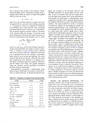
TABLE VII Correlation of Common Proton Chemical Shifts
Group δ Scale τ Scale Nuclear γ -ray resonance spectroscopy. This
technique is based on the resonance absorption of γ ra-
Tetramethylsilane 0 10.00
diation and is more conventionally known as M¨ossbauer
H 3 C C (saturated) 1.3–0.7 8.7–9.3
spectroscopy. The source of the radiation is a nuclide fixed
CH 2 (saturated) 1.5–1.2 8.5–8.8
in a solid crystal lattice held below the Debye tempera-
H 3 C C C 1.9–1.6 8.1–8.4
ture. In this condition, γ radiation of energies less than
H 2 C C C 2.3–1.8 7.7–8.2
150 keV are emitted with no loss of energy. Such quan-
H 3 C Ar 2.5–2.1 7.5–7.9
tized γ photons can undergo resonance absorption by the
H C C (nonconjugated) 2.7–2.4 7.3–7.6
appropriate identical stable nuclide in a solid sample ma-
H C C (conjugated) 3.1–2.8 6.9–7.2
trix. If the chemical environment of the absorbing nu-
H 3 C O 4.0–3.3 6.0–6.7
clide is different from the emitter, energy must be added
C CH (cyclic) 5.7–5.2 4.3–4.8
or subtracted from the radiation to establish resonance.
ArH (benzenoid) 8.0–6.6 2.0–3.4
This can be achieved by introducing net motion to the
R CHO 9.8–9.5 0.2–0.5
source or absorber to establish a Doppler motion energy
R COOH 11.5–11.0 −1.5to −1.0
term.