
Page 101 - Flexible Robotics in Medicine
P. 101
86 Chapter 4
one driving cable while reeling out the opposing driving cable by an equal length. The
tensioned driving cable will cause tilting of the cylindrical segments about the ball bearings
such that the cylindrical segments are brought closer together on the side that this driving
cable is extended through. This thereby induces bending. The cylindrical segments will be
brought further away from each other on the opposite side, which contains the driving cable
that was reeled out. Therefore this antagonistic movement of the gears and cable spools
allows the bending of the flexible tip in a direction mirroring that of the joystick.
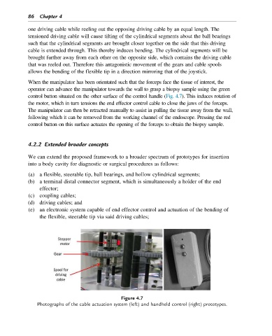
When the manipulator has been orientated such that the forceps face the tissue of interest, the
operator can advance the manipulator towards the wall to grasp a biopsy sample using the green
control button situated on the other surface of the control handle (Fig. 4.7). This induces rotation of
the motor, which in turn tensions the end effector control cable to close the jaws of the forceps.
The manipulator can then be retracted manually to assist in pulling the tissue away from the wall,
following which it can be removed from the working channel of the endoscope. Pressing the red
control button on this surface actuates the opening of the forceps to obtain the biopsy sample.
4.2.2 Extended broader concepts
We can extend the proposed framework to a broader spectrum of prototypes for insertion
into a body cavity for diagnostic or surgical procedures as follows:
(a) a flexible, steerable tip, ball bearings, and hollow cylindrical segments;
(b) a terminal distal connector segment, which is simultaneously a holder of the end
effector;
(c) coupling cables;
(d) driving cables; and
(e) an electronic system capable of end effector control and actuation of the bending of
the flexible, steerable tip via said driving cables;
Figure 4.7
Photographs of the cable actuation system (left) and handheld control (right) prototypes.