
Page 223 - Fundamentals of Light Microscopy and Electronic Imaging
P. 223
206 CONFOCAL LASER SCANNING MICROSCOPY
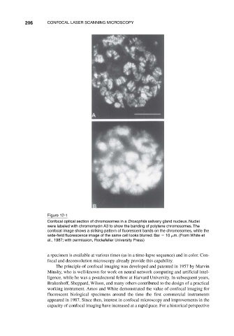
Figure 12-1
Confocal optical section of chromosomes in a Drosophila salivary gland nucleus. Nuclei
were labeled with chromomycin A3 to show the banding of polytene chromosomes. The
confocal image shows a striking pattern of fluorescent bands on the chromosomes, while the
wide-field fluorescence image of the same cell looks blurred. Bar 10 m. (From White et
al., 1987; with permission, Rockefeller University Press)
a specimen is available at various times (as in a time-lapse sequence) and in color. Con-
focal and deconvolution microscopy already provide this capability.
The principle of confocal imaging was developed and patented in 1957 by Marvin
Minsky, who is well-known for work on neural network computing and artificial intel-
ligence, while he was a postdoctoral fellow at Harvard University. In subsequent years,
Brakenhoff, Sheppard, Wilson, and many others contributed to the design of a practical
working instrument. Amos and White demonstrated the value of confocal imaging for
fluorescent biological specimens around the time the first commercial instruments
appeared in 1987. Since then, interest in confocal microscopy and improvements in the
capacity of confocal imaging have increased at a rapid pace. For a historical perspective