
Page 227 - Fundamentals of Light Microscopy and Electronic Imaging
P. 227
210 CONFOCAL LASER SCANNING MICROSCOPY
PMT detector
Pinhole aperture
Dichroic
mirror
Laser
point source
Objective lens
Focal plane
Specimen
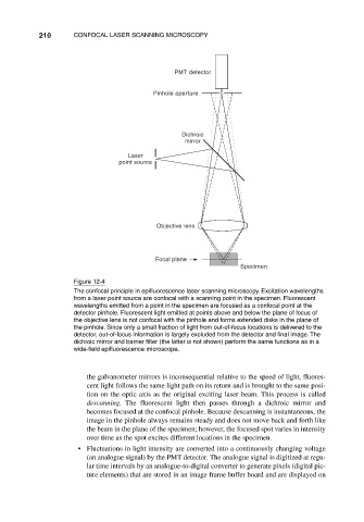
Figure 12-4
The confocal principle in epifluorescence laser scanning microscopy. Excitation wavelengths
from a laser point source are confocal with a scanning point in the specimen. Fluorescent
wavelengths emitted from a point in the specimen are focused as a confocal point at the
detector pinhole. Fluorescent light emitted at points above and below the plane of focus of
the objective lens is not confocal with the pinhole and forms extended disks in the plane of
the pinhole. Since only a small fraction of light from out-of-focus locations is delivered to the
detector, out-of-focus information is largely excluded from the detector and final image. The
dichroic mirror and barrier filter (the latter is not shown) perform the same functions as in a
wide-field epifluorescence microscope.
the galvanometer mirrors is inconsequential relative to the speed of light, fluores-
cent light follows the same light path on its return and is brought to the same posi-
tion on the optic axis as the original exciting laser beam. This process is called
descanning. The fluorescent light then passes through a dichroic mirror and
becomes focused at the confocal pinhole. Because descanning is instantaneous, the
image in the pinhole always remains steady and does not move back and forth like
the beam in the plane of the specimen; however, the focused spot varies in intensity
over time as the spot excites different locations in the specimen.
• Fluctuations in light intensity are converted into a continuously changing voltage
(an analogue signal) by the PMT detector. The analogue signal is digitized at regu-
lar time intervals by an analogue-to-digital converter to generate pixels (digital pic-
ture elements) that are stored in an image frame buffer board and are displayed on