
Page 224 - Fundamentals of Light Microscopy and Electronic Imaging
P. 224
OVERVIEW 207
and annoted bibliography of this technology, refer to articles by Inoué and by Webb in
Pawley (1995).
Confocal microscopes are integrated electronic microscope systems, in which the
light microscope is part of an electronic imaging system containing an electronic detec-
tor or camera, a computer and computer software, and electronic devices for image dis-
play, printing, and storage (Fig. 12-2). The integration is so complete that microscopists
now frequently refer to their microscopes as digital or video imaging systems and their
activity at the microscope as electronic imaging. Electronic microscope systems are rev-
olutionizing the face of research, serving as indispensable tools for documentation and
analysis, and facilitating investigations on molecules, cells, and tissues that until now
have simply not been possible. This transformation is the result of rapid technological
advances in opto-electronics, lasers, fiber optics, thin film dielectric coatings, comput-
ers, printers and image storage devices, and image acquisition and processing software.
In order to use these imaging systems, we must not only learn principles of light
microscopy, but also master electronic imaging and image processing operations. Some
of the challenges involve very fundamental concepts, such as how to take a picture. This
is especially relevant to confocal microscopy, where there are no camera buttons to
push, and even if there were, there is no confocal image to take a picture of! Acquiring
an image with an electronic microscope system requires us to use computer software
and make decisions that affect the resolution of space, time, and light intensity in the
image. Since the ability to make these decisions quickly and with confidence requires
training and education, we focus on electronic image acquisition in this and the follow-
ing chapters.
Scan
head
Computer
Laser
Monitors
Image Menus
Microscope
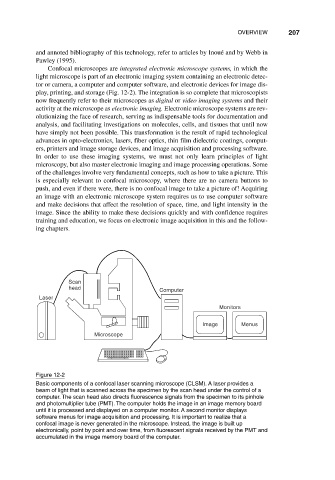
Figure 12-2
Basic components of a confocal laser scanning microscope (CLSM). A laser provides a
beam of light that is scanned across the specimen by the scan head under the control of a
computer. The scan head also directs fluorescence signals from the specimen to its pinhole
and photomultiplier tube (PMT). The computer holds the image in an image memory board
until it is processed and displayed on a computer monitor. A second monitor displays
software menus for image acquisition and processing. It is important to realize that a
confocal image is never generated in the microscope. Instead, the image is built up
electronically, point by point and over time, from fluorescent signals received by the PMT and
accumulated in the image memory board of the computer.