Page 231 - Fundamentals of Light Microscopy and Electronic Imaging
P. 231
214      CONFOCAL LASER SCANNING MICROSCOPY


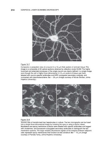
                                Figure 12-7
                                Composite or projection view of a neuron in a 10  m thick section of rat brain tissue. The
                                image is a composite of 20 optical sections obtained by reflection-mode CLSM. The highly
                                branched and extended processes of the single neuron are clearly defined in a single image,
                                even though the cell is highly three-dimensional. A 10  m section of tissue was fixed,
                                labeled with neuron-specific antibodies and HRP-conjugated secondary antibody, and
                                contrasted with diaminobenzidene. Bar   20  m (Image courtesy of Nicole Stricker, Johns
                                Hopkins University.)


                                Figure 12-8
                                Volume view of transformed liver hepatocytes in culture. The two micrographs can be fused
                                into a single three-dimensional image by crossing the eyes or using a stereo viewer.
                                Basilateral and apical membrane surfaces were labeled with specific antibodies and tagged
                                with rhodamine- and fluorescein-conjugated secondary antibodies to distinguish the two
                                membrane systems. The large vesicles (fluorescein signal) at the margins between adjacent
                                cells represent apical membranes that function as bile canaliculi. Bar   10  m (Image
                                courtesy of Pamela Tuma, Johns Hopkins University.)

