
Page 277 - Fundamentals of Light Microscopy and Electronic Imaging
P. 277
260 DIGITAL CCD MICROSCOPY
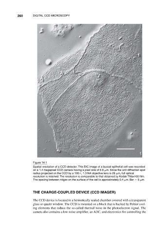
Figure 14-1
Spatial resolution of a CCD detector. This DIC image of a buccal epithelial cell was recorded
on a 1.4 megapixel CCD camera having a pixel size of 6.8 m. Since the unit diffraction spot
radius projected on the CCD by a 100 , 1.3 NA objective lens is 26 m, full optical
resolution is retained. The resolution is comparable to that obtained by Kodak TMax100 film.
The spacing between ridges on the surface of the cell is approximately 0.4 m. Bar 5 m.
THE CHARGE-COUPLED DEVICE (CCD IMAGER)
The CCD device is located in a hermetically sealed chamber covered with a transparent
glass or quartz window. The CCD is mounted on a block that is backed by Peltier cool-
ing elements that reduce the so-called thermal noise in the photoelectron signal. The
camera also contains a low noise amplifier, an ADC, and electronics for controlling the