
Page 373 - Handbook of Properties of Textile and Technical Fibres
P. 373
346 Handbook of Properties of Textile and Technical Fibres
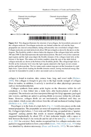
Cleaved via procollagen Cleaved via procollagen
aminoprotease carboxyprotease
150 Å 3000 Å 100 Å
13 67 121 175 229 283 337 391 445 499 553 607 661 715 769 823 877 931 985 1039
Figure 11.1 This diagram illustrates the structure of procollagen, the biosynthetic precursor of
the collagen molecule. Procollagen molecules are formed within the cell and the large
propeptides are cleaved extracellularly during self-assembly into crosslinked collagen fibers.
Collagen molecules are triple helical rods about 300 nm in length as shown by the arrows in the
diagram. The flexibility profile is shown below the diagram of the collagen triple helix and the
300 nm (3000 Å) line that represents the triple helical portion. The dark vertical lines represent
rigid regions and the light areas depict the flexible domains of the collagen triple helix at the
bottom of the figure. The amino acid residue numbers along the axis of the triple helical
collagen molecule are shown at the bottom of the flexibility profile. The collagen triple helix is
flexible at the molecular level since the light regions depicted in the figure do not contain
proline and hydroxyproline. The two imino acids contain rings prevent the formation of a rigid
triple helix. Upon tensile loading the flexible domains stretch, storing mechanical energy that
can later be transmitted to joints to allow movement such as locomotion.
collagen is found in tendons, skin, cornea, bone, lung, and vessel walls (Hulmes,
2008). This collagen is thought to give rise to the high tensile strength of collagen
fibers in tendon; in addition, it actively is involved in other physiologic processes
such as mechanotransduction.
Collagen synthesis from amino acids begins on the ribosomes within the cell
cytoplasm; it is then folded into a triple helix after hydroxylation of proline is
completed. The molecules are then transported through the Golgi apparatus, packaged
into vesicles, and then are added to growing fibrils in recesses within the cell mem-
brane. However, collagen fibrils are not mechanically stabilized until they are
cross-linked, which occurs after release from the cell and mechanical loading begins
(McBride et al., 1985, 1988).
Procollagen is in the form of a triple helix (Fig. 11.1) with extra pieces on the ends
termed propeptides. The propeptides are removed sequentially during collagen assem-
bly. Collagen triple helices, 1.5 nm wide and 300 nm long, are packed into a “quarter-
staggered” packing pattern that results in nearest neighboring molecules being
staggered longitudinally by about 22% of their molecular lengths with a space or
hole between the head of one molecule and the tail of the next (Fig. 11.2) (see Silver
et al., 2003 for a review). Early studies recognized that five collagen molecules were
packed in staggered fashion to form a microfibril (Smith, 1968; Doyle et al., 1974) that
was identified by electron microscopy (Pease and Bouteille, 1972). We now believe