
Page 374 - Handbook of Properties of Textile and Technical Fibres
P. 374
Structure and behavior of collagen fibers 347
Overlap region Hole region
D D D
Overlap region Hole region
c2 c1 b2 b1 a4 a3 a2 a1 e2 e1 d c3
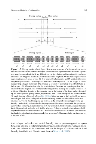
Figure 11.2 The top portion of this figure illustrates the structure of a five-membered micro-
fibrillar unit that is believed to be the repeat unit found in collagen fibrils and fibers and appears
as a quasi-hexagonal unit by X-ray diffraction of tendon. In this packing pattern five collagen
molecules are staggered by about 22% of the molecular length of 300 nm with respect to their
nearest neighbors. A space or hole 0.6 D in length (D is between 64 and 67 nm) is left between
neighboring molecules. The collagen molecule is 4.4 D long where D is the stagger between
neighboring collagen molecules. The distance D consists of an overlap zone of 0.4 D and a
hole region of 0.6 D as is shown by the vertical dotted lines that are superimposed on the
microfibril in the diagram. The overlap and hole regions that make up the D repeat consist of 13
rigid and 12 flexible domains in the expanded view at the bottom of the figure and are depicted
by the rectangles and springs shown, respectively. The 12 flexible regions are identical to the
12 bands denoted c2 through c3 (see Fig. 11.3(d,e)) that are seen as dark vertical lines across
the collagen fibril when collagen is stained with heavy metals and viewed in the electron
microscope. The 12 flexible regions are believed to be stretched when collagen fibrils are
initially mechanically deformed reflecting experimental increases in the axial rise per amino
acid residue, h spacing, that are observed by X-ray diffraction. Further loading causes increases
in the D period and molecular and fibrillar slippage. Collagen molecules in tendon are held
together in the microfibril with crosslinks that occur at the tail of one molecule (see circle)to
the head of a lateral neighboring molecule (see arrowhead). These crosslinks are staggered by
a distance of 4D.
that collagen molecules are packed laterally into a quarter-staggered or quasi-
hexagonal unit that is in turn longitudinally packed into microfibrils. Collagen micro-
fibrils are believed to be continuous and run the length of a tissue and are fused
laterally into fibrils and fibers in most tissues (Silver et al., 2003).