
Page 82 - Handbook of Properties of Textile and Technical Fibres
P. 82
Properties of wool 63
R O R O R O R O
H H H H
N N N N
HN N N N N
H H H H
HN O CH O CH 2 O CH 2 O CH 2
O H 3 C CH 3 H 2 C O OC
Hydrophobic bond H
CH 2 Hydrogen NH
CH 2
bond Isopeptide
Intermolecular S H 2 C Ionic O H 2 C cross-link
disulfide cross-link bond NH 2
S NH 3 C CH 2
COO H 2 C H 2 C
CH 2
HN O CH 2 O H 2 C O CH 2 O CH 2
H H H H H
N N N N N
N N N N
O H H H H
R O H 2 C O CH 2 O R O
S S
Intramolecular
disulfide cross-link
Figure 3.3 Types of covalent and noncovalent bonds in wool (Rippon et al., 2016).
3.2.2 Physical
The wool fiber has a complex hierarchical structure, which is shown schematically in
Fig. 3.5. As already mentioned the low-sulfur proteins can form a-helical structures
that assemble into rodlike intermediate filaments (microfibrils). Two right-handed
a-helices twist together to form a left-handed coiled-coil structure. Four of these dou-
ble helix structures in turn self-assemble to form a protofilament of about 2 nm in
diameter and finally eight protofilaments form a ring-type arrangement to generate a
microfibril (Wortmann and Zahn, 1994) with a diameter of about 7 nm and a length
of at least 1 mm(Rippon, 1992). The crystalline microfibrils are embedded in an amor-
phous cross-linked matrix (Fig. 3.6), with many microfibrils grouped together to form
15
Radial swelling (%) 10 5
0
0 20 40 60 80 100
Relative humidity (%)
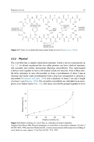
Figure 3.4 Radial swelling of a wool fiber as a function of relative humidity.
Adapted from Huson MG: Physical properties of wool fibres in electrolyte solutions, Text Res J
68:595e605, 1998; data from Warburton FL: A direct measurement of the transverse swelling of
wool fibres in water vapour, J Text Inst 38:T65eT72, 1947.