
Page 85 - Handbook of Properties of Textile and Technical Fibres
P. 85
66 Handbook of Properties of Textile and Technical Fibres
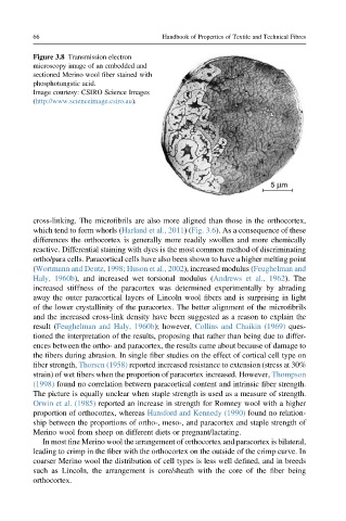
Figure 3.8 Transmission electron
microscopy image of an embedded and
sectioned Merino wool fiber stained with
phosphotungstic acid.
Image courtesy: CSIRO Science Images
(http://www.scienceimage.csiro.au).
cross-linking. The microfibrils are also more aligned than those in the orthocortex,
which tend to form whorls (Harland et al., 2011)(Fig. 3.6). As a consequence of these
differences the orthocortex is generally more readily swollen and more chemically
reactive. Differential staining with dyes is the most common method of discriminating
ortho/para cells. Paracortical cells have also been shown to have a higher melting point
(Wortmann and Deutz, 1998; Huson et al., 2002), increased modulus (Feughelman and
Haly, 1960b), and increased wet torsional modulus (Andrews et al., 1962). The
increased stiffness of the paracortex was determined experimentally by abrading
away the outer paracortical layers of Lincoln wool fibers and is surprising in light
of the lower crystallinity of the paracortex. The better alignment of the microfibrils
and the increased cross-link density have been suggested as a reason to explain the
result (Feughelman and Haly, 1960b); however, Collins and Chaikin (1969) ques-
tioned the interpretation of the results, proposing that rather than being due to differ-
ences between the ortho- and paracortex, the results came about because of damage to
the fibers during abrasion. In single fiber studies on the effect of cortical cell type on
fiber strength, Thorsen (1958) reported increased resistance to extension (stress at 30%
strain) of wet fibers when the proportion of paracortex increased. However, Thompson
(1998) found no correlation between paracortical content and intrinsic fiber strength.
The picture is equally unclear when staple strength is used as a measure of strength.
Orwin et al. (1985) reported an increase in strength for Romney wool with a higher
proportion of orthocortex, whereas Hansford and Kennedy (1990) found no relation-
ship between the proportions of ortho-, meso-, and paracortex and staple strength of
Merino wool from sheep on different diets or pregnant/lactating.
In most fine Merino wool the arrangement of orthocortex and paracortex is bilateral,
leading to crimp in the fiber with the orthocortex on the outside of the crimp curve. In
coarser Merino wool the distribution of cell types is less well defined, and in breeds
such as Lincoln, the arrangement is core/sheath with the core of the fiber being
orthocortex.