
Page 84 - Handbook of Properties of Textile and Technical Fibres
P. 84
Properties of wool 65
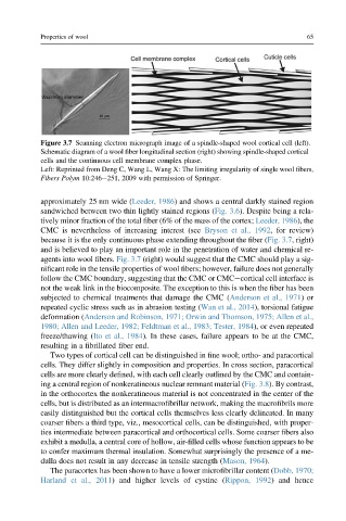
Figure 3.7 Scanning electron micrograph image of a spindle-shaped wool cortical cell (left).
Schematic diagram of a wool fiber longitudinal section (right) showing spindle-shaped cortical
cells and the continuous cell membrane complex phase.
Left: Reprinted from Deng C, Wang L, Wang X: The limiting irregularity of single wool fibers,
Fibers Polym 10:246e251, 2009 with permission of Springer.
approximately 25 nm wide (Leeder, 1986) and shows a central darkly stained region
sandwiched between two thin lightly stained regions (Fig. 3.6). Despite being a rela-
tively minor fraction of the total fiber (6% of the mass of the cortex; Leeder, 1986), the
CMC is nevertheless of increasing interest (see Bryson et al., 1992, for review)
because it is the only continuous phase extending throughout the fiber (Fig. 3.7, right)
and is believed to play an important role in the penetration of water and chemical re-
agents into wool fibers. Fig. 3.7 (right) would suggest that the CMC should play a sig-
nificant role in the tensile properties of wool fibers; however, failure does not generally
follow the CMC boundary, suggesting that the CMC or CMCecortical cell interface is
not the weak link in the biocomposite. The exception to this is when the fiber has been
subjected to chemical treatments that damage the CMC (Anderson et al., 1971)or
repeated cyclic stress such as in abrasion testing (Wan et al., 2014), torsional fatigue
deformation (Anderson and Robinson, 1971; Orwin and Thomson, 1975; Allen et al.,
1980; Allen and Leeder, 1982; Feldtman et al., 1983; Tester, 1984), or even repeated
freeze/thawing (Ito et al., 1984). In these cases, failure appears to be at the CMC,
resulting in a fibrillated fiber end.
Two types of cortical cell can be distinguished in fine wool; ortho- and paracortical
cells. They differ slightly in composition and properties. In cross section, paracortical
cells are more clearly defined, with each cell clearly outlined by the CMC and contain-
ing a central region of nonkeratineous nuclear remnant material (Fig. 3.8). By contrast,
in the orthocortex the nonkeratineous material is not concentrated in the center of the
cells, but is distributed as an intermacrofibrillar network, making the macrofibrils more
easily distinguished but the cortical cells themselves less clearly delineated. In many
coarser fibers a third type, viz., mesocortical cells, can be distinguished, with proper-
ties intermediate between paracortical and orthocortical cells. Some coarser fibers also
exhibit a medulla, a central core of hollow, air-filled cells whose function appears to be
to confer maximum thermal insulation. Somewhat surprisingly the presence of a me-
dulla does not result in any decrease in tensile strength (Mason, 1964).
The paracortex has been shown to have a lower microfibrillar content (Dobb, 1970;
Harland et al., 2011) and higher levels of cystine (Rippon, 1992) and hence