
Page 179 - Inorganic Mass Spectrometry - Fundamentals and Applications
P. 179
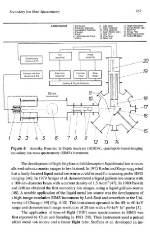
Secondary Ion  Mass  Spectrometry                             167


                              A-DIDA System   1  ion  Source   8  Gate  Valve  15  Target  Manipulator
                                         2 Extraction  Electrode   9  Beam  Deflection  16  Sam   le Holder  (opt.)
                                         3 Acceleration  Electrode  10  Orfice  (optlonal)  17  Blana)  Po?
                                         4  Insulator   11  Beam  Monitor   (opt.)   18 Energy  Filter
                                         5  Cover     12 View  Port  19  Mass  Spectrometer
                                         6 Turbomolecular  Pump   13 UHV-Pump   20  Particle  Multiplier
                                         7  Orifice   14 Blank  Port


                   Atomika  Dynamic In Depth  Analyzer  (ADIDA), quadrupole-based-imaging
          secondary ion mass  spectrometry  (SIMS) instrument.

               The development of high-brightness  field  desorption  liquid  metal  ion  sources
          allowed sub~crometer images  to be obtained.  In  1975  Krohn  and  Ringo  suggested
          that a finely  focused  liquid  metal  ion  source  could be used for scanning  probe  SIMS
                                  et
          imaging  [46].  In  1979  Seliger al.  demonstrated a liquid  gallium  ion  source  with
                                  a
          a 100-nm-diameter  beam  with current  density of 1.5 Ncm2 [47].  In  1980  Prewitt
          and  Jeffries  obtained the first  secondary  ion  images,  using liquid  gallium  source
                                                         a
                                                                         of
          E4.81. A notable  application of the  liquid  metal  ion  source  was  the  development
          a high-image-resolution  SIMS  instrument by Levi-Setti  and  coworkers  at the Uni-
          versity of Chicago  [49]  (Fig.  4.10). This instrument  operated  in  the  4.0- to 60-keV
          range and  demonstrated  image  resolution  of 20 nm  with a 4.0-keV  In* probe  [3].
               The application of  time-of-flight (TOE;) mass  spectrometers to SMS was
          first  reported by Chait and  Standing  in 198  1 [so]. Their  instrument  used a pulsed
          alkali metal  ion  source  and a linear  flight  tube.  Steffens et al.  developed an im-