
Page 180 - Inorganic Mass Spectrometry - Fundamentals and Applications
P. 180
Cristy
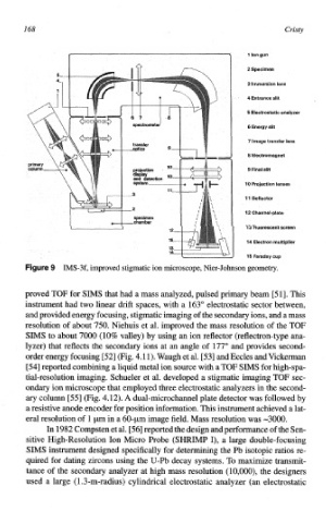
f 1 Ion gun
2 Specimen
3 Immersion lens
4 Entrance slit
S Electrostatic analyzer
6 Energy slit
7 Image transfer lens
8 ~lectromagnet
9 Final slit
10 Projection lensas
11 Deflector
12 Chann~l-plate
13 Fluorescent screen
14 Electron multiplier
15 Faraday cup
ure 9 IMS-3f, improved Stigmatic ion microscope, Nier-Johnson geometry.
proved TOF for SIMS that had a mass analyzed, pulsed primary beam [51]. This
inst~ment had two linear drift spaces, with a 163" electrostatic sector between,
and provided energy focusing, stigmatic imaging of the secondary ions, and a mass
resolution of about 750. Niehuis et al. improved the mass resolution of the TOF
SIMS to about 7000 (10% valley) by using an ion reflector (reflectron-type ana-
lyzer) that reflects the secondary ions at an angle of 177" and provides second-
order energy focusing [52] (Fig. 4.1 1). Waugh al. [53] and Eccles and Viclceman
et
[54] reported combining a liquid metal ion source with a TOF SIMS for high-spa-
tial-resolution imaging. Schueler et al. developed a stigmatic imaging TOF sec-
ondary ion microscope that employed thee electrostatic analyzers in the second-
ary colum E551 (Fig. 4.12). A dual-~crochannel plate detector was followed by
a resistive anode encoder for position infomation. This ins~~ment achieved a lat-
eral resolution of 1 pm in a 60-pm image field. Mass resolution was -3000,
In 1982 Compsten et al. [56] reported the design and performance of the Sen-
sitive High-Resolution Ion Micro Probe (SHRIMP I), a large double-focusing
SIMS ins~ment designed specifically for dete~ning the Pb isotopic ratios re-
quired for dating zircons using the U-Pb decay systems. To maximize transmit-
tance of the secondary analyzer at high mass resolution (lO,OOO), the designers
used a large (l .3-m-radius) cylindrical electrostatic analyzer (an electrostatic