
Page 222 - Inorganic Mass Spectrometry - Fundamentals and Applications
P. 222
Seconda~ Ion Mass Spectrometry 207
'" l
'IO'F;) 50 100 150 200 250 300
Depth (A)
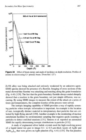
Effect of beam energy and angle of incidence on depth resolution. Profiles of
arsenic in silicon using CS' primary beam. (From Ref. 127.)
Zr-Nb alloy was being attacked and seriously weakened by an unknown agent.
SMS spectra showed the presence of a fluoride. Imaging of cross sections of the
metal showed that fluorine was attacking and reacting along the grain boundaries
(Fig. 4.40) [129]. The fact that the grain boundary fluoride always ended abruptly
showed that a reaction in the grain boundary, not just simple difTusion, was oc-
curring. By using SIMS images to measure the extent of the attack at a series of
times and temperatures, the complete kinetics of the process were solved.
The isotopic imaging capability of SIMS provides a way of rapidly screen-
ing particles when isotopic info~ation is important. An example is the location
of rare, isotopically distinct material in inte~lanetary dust particles that are col-
lected by high-flying aircraft [130]. Another example the monito~ng of nuclear
is
enrichment facilities by environme~tal sampling that requires quick scanning of
particles to detect enriched uranium [ 13 l]. Sirnons et al. reported an automated
SIMS for rapidly determining isotopic distributions in particles [ 1321.
A study of engineered silver halide crystals used the high resolving power
of a liquid metal ion gun to image 0.1- to 0.5-pm-thick layers of AgBr and
AgBr,,I,, that were grown on AgBr platelets (Fig.
4.41) [ 1331. The flat platelets