
Page 225 - Inorganic Mass Spectrometry - Fundamentals and Applications
P. 225
21 0
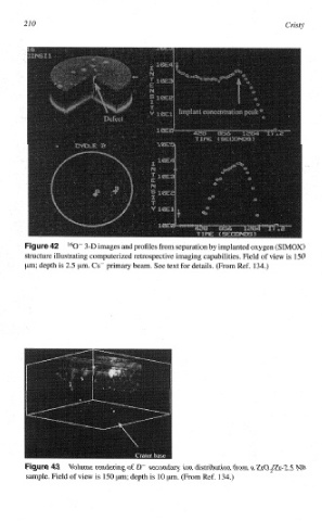
l60" 3-D images and profiles from separation by implanted oxygen (SIMOS)
structure illustrating computerized retrospective imaging capabilities. Field of view is 150
pm; depth is 2.5 pm. CS- primary beam. See text for details. (From Ref. 134.)
Volume rendering of D- secondary ion distribution from a Zr02/2r-2.5 I%
sample. Field of view is I50 pm; depth is 10 pm. (From Ref. 134.)