
Page 292 - Optofluidics Fundamentals, Devices, and Applications
P. 292
266 Cha pte r Ele v e n
4
1
r
Point source 3 Transmission (a.u.) 0.5
Z Z
H Resolution of OFM (μm) 2 0 –5 0 5
Y r (μm)
X
Al 1
SU8
CMOS pixel 0
10 –2 10 –1 10 0 10 1
H (μm)
(b)
(a)
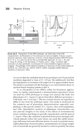
FIGURE 11-4 Resolution of the OFM prototype. (a) Schematic of the PSF
measurement. (b) Resolution of the prototype at various heights H above a 1-μm-
diameter aperture under Sparrow’s criterion. (Inset) Representative OFM PSF plots
at H = 0.1, 1.5, and 2.5 μm for the 1-μm-diameter aperture. (X. Cui, L. M. Lee,
X. Heng, W. Zhong, P. W. Sternberg, D. Psaltis, and C. Yang, “Lensless high-
resolution on-chip optofl uidic microscopes for Caenorhabditis elegans and cell
imaging,” Proceedings of the National Academy of Sciences of the United States of
America, vol. 105 (31), pp. 10670–10675, 2008. Copyright (2008) National
Academy of Sciences, USA.)
we can see that the resolution limit of our prototype was 0.9 μm and the
resolution degraded to 3 μm at H = 2.5 μm. We additionally find that
the degradation of resolution with respect to H is approximately linear.
The result is consistent with the findings in our more detailed study on
aperture-based imaging systems in Ref. 6.
As an illustration of the OFM’s utility for bioscience applica-
tions, we further implemented a demonstration experiment where
we used the OFM prototype to image and analyze the body size
phenotypes of C. elegans L1 larvae from three different genotypes—
wild-type, sma-3, and dpy-7. The two mutant strains are expected to
differ in size from the wild-type strain. This study is motivated by
the extensive use of phenotype characterization, especially mor-
phology, in the genetic studies of C. elegans, other microorganisms,
and cells. Quantitative analysis of phenotype characteristics can
also provide insights in the study of the effects of environmental
stress, nutrition and drugs on growth, reproduction, and life span of
C. elegans. Often, the variation in physical dimensions amongst indi-
vidual animals within the same strain can mask the subtle pheno-
type difference between different strains. As such, to perform mean-
ingful phenotype characterization, measurements on a large sample
size of nematode are generally needed in order to quantify the mean