
Page 444 - Polymer-based Nanocomposites for Energy and Environmental Applications
P. 444
Development of polymer nanocomposites 401
well diffused uniformly with increasing concentrations of silver nitrate. The 5 mM
silver nanocomposite film is dark brown, revealing that uniform distribution of AgNPs
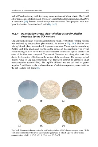
in the matrix [19]. Further, the cellulose/silver-nanocoated films prepared were ana-
lyzed for biofilm formation by E. coli (Fig. 14.3).
14.3.4 Quantitative crystal violet-binding assay for biofilm
detection by the TCP method
The antifouling efficacy of silver nanocomposite with E. coli biofilm-forming bacteria
was analyzed by tissue-culture plate method. As shown in Fig. 14.4, biofilm con-
taining 24-well plate, it treated with Ag nanocomposites. The composites containing
AgNPs inhibit the attachment biofilm on the surface of the membrane. The crystal
violet binding cells of silver treated and untreated composites optical density and
color of the film were compared. The control film color was changed to dark blue
due to the formation of biofilm on the surface of the membrane. The average optical
density value of Ag nanocomposites was decreased contrast to untreated silver
nanocomposites (control film). The AgNPs diffused into the cell wall of gram-
negative E. coli bacteria, the vital constituents of cellular components, come out from
the cell leads to cell death [1].
(A) (B) (C)
(D) (E) (F)
Fig. 14.3 Silver-coated composites for antifouling studies: (A) Cellulose composite and (B–F)
cellulose composites with silver nanoparticles generated in situ at aqueous silver nitrate
concentrations of (B) 1, (C) 2, (D) 3, (E) 4, and (F) 5 mM.