
Page 446 - Polymer-based Nanocomposites for Energy and Environmental Applications
P. 446
Development of polymer nanocomposites 403
attachment of biofilm was drastically reduced the increasing concentration of silver
nanoparticle. The 5 mM concentration of antifouling sample biofilm attachment
was only 6.9%, and the inhibition of biofilm was 93.1%. The 1 mM concentration
of antifouling sample biofilm attachment was 36.79%, and the calculated biofilm inhi-
bition was 63.21%. The 3 mM concentration of antifouling sample biofilm inhibition
was 84.48%, and the calculated biofilm attachment was 15.52% [20].
14.3.6 SEM analysis of antifouling samples
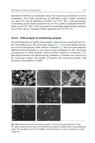
The antifouling effect of AgNP-coated polymer composites was evaluated by the crys-
tal violet binding assay. The microscopic image Fig. 14.6A reveals uniform distribu-
tion of silver nanoparticles in the cellulose composites [1]. The silver nanocomposite
inhibited biofilm formation, as was observed after incubation for 48 h. Increasing
concentrations of AgNP markedly reduced biofilm formation in composites [20].
Rod-shaped bacteria with interconnecting formations of biofilm were observed in
the microscopic images. The number of bacterial cells decreased gradually with
increasing concentrations of AgNP.
Fig. 14.6 Scanning electron microscopic images of silver-nanocoated polymeric films:
(A) AgNP-coated cellulose film, (B–D) biofilm developed on cellulose/silver nanocomposites
during 48 h incubation, and (E–H) biofilm reduction efficacy of silver nanoparticles on the
composites.