
Page 144 - Vibrational Spectroscopic Imaging for Biomedical Applications
P. 144
120 Cha pte r F o u r
(a) (b)
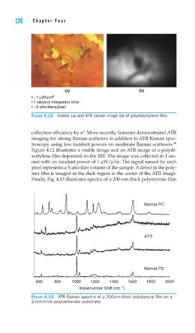
• ~1 μW/μm 2
• 1 second integration time
• ~5 atto-liters/pixel
FIGURE 4.12 Visible (a) and ATR raman image (b) of polydiacetylene fi lm.
2
collection efficiency by n . More recently, Sommer demonstrated ATR
imaging for strong Raman scatterers in addition to ATR Raman spec-
troscopy using low incident powers on moderate Raman scatterers. 56
Figure 4.12 illustrates a visible image and an ATR image of a polydi-
acetylene film deposited on the IRE. The image was collected in 1 sec-
2
ond with an incident power of 1 μW/μ m. The signal sensed by each
pixel represents a 5-atto-liter volume of the sample. A defect in the poly-
mer film is imaged as the dark region in the center of the ATR image.
Finally, Fig. 4.13 illustrates spectra of a 200-nm-thick polystyrene film
Normal PC
ATR
Normal PS
600 800 1000 1200 1400 1600 1800 2000
–1
Wavenumber Shift (cm )
FIGURE 4.13 ATR Raman spectra of a 200-nm-thick polystyrene fi lm on a
2-mm-thick polycarbonate substrate.