
Page 355 - Vibrational Spectroscopic Imaging for Biomedical Applications
P. 355
T issue Imaging with CARS Micr oscopy 329
1.2
Relative CARS Intensity 0.8
1.0
0.6
0.4
0.2
0.0
2800 2900 3000 3100 3200 3300 3400 3500
–1
Wavenumber (cm )
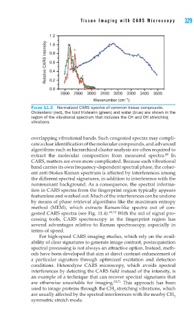
FIGURE 11.3 Normalized CARS spectra of common tissue compounds.
Cholesterol (red), the lipid tristearin (green) and water (blue) are shown in the
region of the vibrational spectrum that includes the CH and OH stretching
vibrations.
overlapping vibrational bands. Such congested spectra may compli-
cate a clear identification of the molecular compounds, and advanced
algorithms such as hierarchical cluster analysis are often required to
68
extract the molecular composition from measured spectra. In
CARS, matters are even more complicated. Because each vibrational
band carries its own frequency-dependent spectral phase, the coher-
ent anti-Stokes Raman spectrum is affected by interferences among
the different spectral signatures, in addition to interference with the
nonresonant background. As a consequence, the spectral informa-
tion in CARS spectra from the fingerprint region typically appears
featureless and washed out. Much of the interferences can be undone
by means of phase retrieval algorithms like the maximum entropy
method (MEM), which extracts Raman-like spectra out of con-
gested CARS spectra (see Fig. 11.4). 69,70 With the aid of signal pro-
cessing tools, CARS spectroscopy in the fingerprint region has
several advantages relative to Raman spectroscopy, especially in
terms of speed.
For high-speed CARS imaging studies, which rely on the avail-
ability of clear signatures to generate image contrast, postacquisition
spectral processing is not always an attractive option. Instead, meth-
ods have been developed that aim at direct contrast enhancement of
a particular signature through optimized excitation and detection
conditions. Heterodyne CARS microscopy, which avoids spectral
interferences by detecting the CARS field instead of the intensity, is
an example of a technique that can recover spectral signatures that
are otherwise unsuitable for imaging. 53,71 This approach has been
used to image proteins through the CH stretching vibrations, which
3
are usually affected by the spectral interferences with the nearby CH
2
symmetric stretch mode.