
Page 360 - Vibrational Spectroscopic Imaging for Biomedical Applications
P. 360
334 Cha pte r Ele v e n
increase the number of in-phase photons that arrive in the focal vol-
ume at greater depths, higher excitation powers can be used. Such
an approach has been used to accomplish deep-tissue imaging with
91
two-photon-excited fluorescence, and is, in principle, also possible
for CARS microscopy. Absorption and linear heating of the sample are
the limiting factors for applying more power to achieve strong signals
at greater depths. The photodamage threshold in pigmented skin, for
2 92
instance, is found to be 500 W/cm . Generally, when keeping sample
illumination dosages below 50 mW per beam, imaging well below
the damage threshold can be achieved.
Besides lower signals as a consequence of random scattering
throughout the tissue, scattering also affects the CARS imaging prop-
erties when scattering objects in focus affect the signal generation
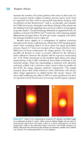
process. Figure 11.7 shows an example of how linear refractive index
differences in focus compromise the image quality. The image of a
paraffin oil droplet in water is severely affected by the refractive
index difference between the droplet and its aqueous surrounding
Δn ~.015. Phase distortions of the incident light along with phase
mismatching of the CARS radiation in focus both contribute to the
distorted image. When the surrounding is replaced with dimethyl
sulfoxide, a fluid with a refractive index closer to that of paraffin oil
Δn = 003. , the image appears relatively undistorted. This simple
example shows that linear refractive index differences will always
affect image appearance in turbid media like tissues. Figure 11.8
shows that scattering may affect CARS in a more significant way than
two-photon-excited fluorescence microscopy. In this regard, a proper
(a) (b)
(1)
(3)
FIGURE 11.7 Effect of χ scattering on coherent χ signals. (a) CARS image
of a dodecane droplet in water. Clear vertical shadow edges can be seen in
nonresonant signal from the water, which is a direct consequence of linear
scattering of light at the dodecane/water interfaces. (b) Paraffi n droplet in d-
DMSO. Because the refractive index differences between paraffi n and d-DMSO
are minimal, the shadowing effect is much reduced.