
Page 363 - Vibrational Spectroscopic Imaging for Biomedical Applications
P. 363
T issue Imaging with CARS Micr oscopy 337
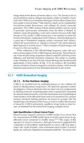
image taken in the dermis of human skin ex vivo. The dermis is rich in
structural fibers such as collagen and elastin, which can both be visual-
ized with CARS, as is evident from the figure. Similar observations have
84
been made in arterial tissue. Alternatively, elastin can be visualized by
two-photon-excited fluorescence and collagen by second harmonic
generation. The CARS contrast of these structural fibers may be useful
if molecular spectroscopic information from the fibers is desired. In
addition, tissue fat generates a very clear contrast, because of the high
density of CH modes. CARS microscopy is the method of choice for
2
studies that require visualization of fat in tissues, which has been put to
a good use in biomedical imaging studies concerned with obesity-
98
related fat accumulation in mammary tissues and atherosclerotic
65
lipid deposits in arterial tissue. More examples of lipid images will
be given in the next section.
When addressed at the OH-stretching frequency, water also pro-
duces strong signals in the CARS imaging microscope. Visualizing tis-
sue water at rapid image acquisition times is useful for following water
diffusion and real-time hydration dynamics. The ability to monitor
water diffusion is not only relevant to tissue biology, but has also found
applications in food science. In Fig. 11.11, for instance, the hydration
process of water in cheese is mapped as a function of time, which reveals
important information on how hydration depends on fat content.
11.7 CARS Biomedical Imaging
11.7.1 Ex Vivo Nonlinear Imaging
CARS is an excellent tool for examining tissues ex vivo without the
need for labeling tissue components. The label-free approach enables
investigation of tissue structures that are intact and not compromised
by labeling protocols. Examining intact tissue is particularly important
for disease-related research, where the biochemical and morphological
characteristics of the diseased tissue need to be preserved for a proper
analysis. Standard staining protocols are known to severely alter the
morphology and integrity of the tissue, as well as to affect the presence
of tissue fat. CARS is particularly suitable to image lipids in intact tis-
sues, as illustrated by the biomedical imaging examples below.
Lipid Quantification in Breast Cancer Tissue
Recent nuclear magnetic resonance (NMR) studies have shown that
the concentration of NMR-visible lipids in breast cancer tissue is sig-
nificantly lower compared to healthy tissue. 99,100 The origin of this
signature of cancer is unknown, although it has been suggested that
a depletion of intracellular lipid droplets in cancer cells may play a
major role. Lipid droplets, (sub)-micrometer-sized bodies of neutral
14
lipids, are a natural component of mammary epithelia. In cancer cells,