
Page 367 - Vibrational Spectroscopic Imaging for Biomedical Applications
P. 367
T issue Imaging with CARS Micr oscopy 341
images. In combination with two-photon-excited fluorescence con-
trast of elastin and SHG contrast of collagen, a comprehensive map-
ping of the major arterial tissue components can be accomplished. 65
Such three-dimensional chemical maps of intact arterial tissue are
invaluable for a better understanding of atherosclerotic lesion devel-
opment as a function of kidney function and diet.
11.7.2 In Vivo Nonlinear Imaging
For in vivo imaging applications, the issue of photodamage is par-
ticularly relevant. To limit tissue illumination in a particular location,
fast scanning is imperative. When imaging fat in superficial tissue
layers, the CARS signals are generally high enough to allow for video-
rate imaging. At these rapid image acquisition times, localized tissue
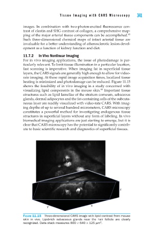
heating is minimized and photodamage can be reduced. Figure 11.15
shows the feasibility of in vivo imaging in a study concerned with
95
visualizing lipid components in the mouse skin. Important tissue
structures such as lipid lamellae of the stratum corneum, sebaceous
glands, dermal adipocytes and the fat-containing cells of the subcuta-
neous layer are readily visualized with video-rate CARS. With imag-
ing depths of up to several hundred micrometers, CARS microscopy
constitutes a powerful method for investigating endogenous tissue
structures in superficial layers without any form of labeling. In vivo
biomedical imaging applications are just starting to emerge, but it is
clear that CARS microscopy has the potential to significantly contrib-
ute to basic scientific research and diagnostics of superficial tissues.
z
y x
FIGURE 11.15 Three-dimensional CARS image with lipid contrast from mouse
skin in vivo. Lipid-rich sebaceous glands near the hair follicle are clearly
3
recognized. Data stack measures 800 × 640 × 125 μm .