
Page 366 - Vibrational Spectroscopic Imaging for Biomedical Applications
P. 366
340 Cha pte r Ele v e n
Lipids in Atherosclerotic Lesions
Atherosclerosis is a disease that affects the walls of arterial blood ves-
sels, forming pools of lipid-rich macrophages, smooth muscle cells,
lipids, and components of the extracellular matrix. These lesions
develop a fibrous encapsulation, which can become increasingly thin
and may rupture as the lesion matures. Rupture of the fibrous cap
causes the release of the inflammatory elements into the lumen,
which, in turn, may obstruct blood flow. 104,105 Multimodal CARS
microscopy is ideally suited to characterize the different stages of the
lesion. A CARS-based methodology may eventually grow into a fiber-
based diagnostic tool for early diagnosis of this arterial disease. Several
studies on carotid arteries of Yorkshire pigs have underlined the
potential of CARS microscopy for atherosclerosis research. 65,84 Here
we present the results of an imaging study on a mouse model system.
The advantage of the ApoE-deficient mouse model is that it enables
the study of atherosclerotic plaques as a function of multiple control-
lable parameters.
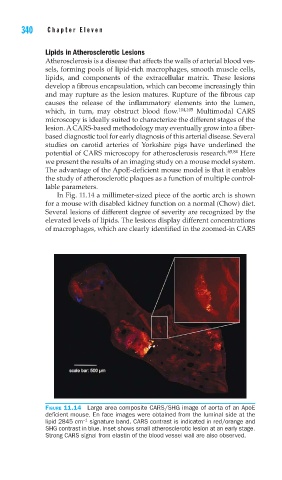
In Fig. 11.14 a millimeter-sized piece of the aortic arch is shown
for a mouse with disabled kidney function on a normal (Chow) diet.
Several lesions of different degree of severity are recognized by the
elevated levels of lipids. The lesions display different concentrations
of macrophages, which are clearly identified in the zoomed-in CARS
FIGURE 11.14 Large area composite CARS/SHG image of aorta of an ApoE
defi cient mouse. En face images were obtained from the luminal side at the
−1
lipid 2845 cm signature band. CARS contrast is indicated in red/orange and
SHG contrast in blue. Inset shows small atherosclerotic lesion at an early stage.
Strong CARS signal from elastin of the blood vessel wall are also observed.