
Page 362 - Vibrational Spectroscopic Imaging for Biomedical Applications
P. 362
336 Cha pte r Ele v e n
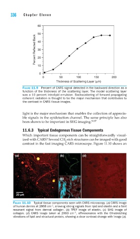
60
Percent Reflected Back 50
40
30
20
10
0
0 50 100 150 200
Thickness of Scattering Layer (μm)
FIGURE 11.9 Percent of CARS signal detected in the backward direction as a
function of the thickness of the scattering layer. The model scattering layer
was a 10 percent intralipid emulsion. Backscattering of forward propagating
coherent radiation is thought to be the major mechanism that contributes to
the contrast in CARS tissue images.
light is the major mechanism that enables the collection of apprecia-
ble signals in the epidetection channel. The same principle has also
been shown to be important in SHG imaging. 96,97
11.6.3 Typical Endogenous Tissue Components
Which important tissue components can be straightforwardly visual-
ized with CARS? Several CH -rich structures can be imaged with good
2
contrast in the fast imaging CARS microscope. Figure 11.10 shows an
(a) (b) (d)
(c)
FIGURE 11.10 Typical tissue components seen with CARS microscopy. (a) CARS image
−1
of human dermis at 2868 cm , showing strong signals from lipid and elastin and a faint
resonant signal from dermal collagen. (b) TPEF image of elastin. (c) SHG image of
−1
collagen. (d) CARS image taken at 2993 cm , off-resonance with the CH-stretching
vibrations of lipid and structural protein, showing a clear contrast change with image (a).