
Page 361 - Vibrational Spectroscopic Imaging for Biomedical Applications
P. 361
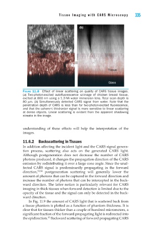
T issue Imaging with CARS Micr oscopy   335


            (a)                        (b)


                                                       Tissue


                                                       Glass

        FIGURE 11.8  Effect of linear scattering on quality of CARS tissue images.
        (a) Two-photon-excited autofl uorescence  xz-image of chicken breast tissue,
        excited at 800 nm using a 1.2-NA water immersion lens. Total scan depth is
        80 μm. (b) Simultaneously detected CARS signal from water. Note that the
        penetration depth of CARS is less than for two-photon-excited fl uorescence,
        and that the coherent third-order signal is more sensitive to linear scattering
        at dense objects. Linear scattering is evident from the apparent shadowing
        streaks in the image.


        understanding of these effects will help the interpretation of the
        images.

        11.6.2 Backscattering in Tissues
        In addition affecting the incident light and the CARS signal genera-
        tion process, scattering also acts on the generated CARS light.
        Although postgeneration does not decrease the number of CARS
        photons produced, it changes the propagation direction of the CARS
        emission by redistributing it over a large cone angle. Since the unaf-
        fected CARS signal is predominantly propagating in the forward
        direction, 93,94  postgeneration scattering will generally lower the
        amount of photons that can be captured in the forward direction and
        increase the number of photons that can be intercepted in the back-
        ward direction. The latter notion is particularly relevant for CARS
        imaging in thick tissues when forward detection is limited due to the
        opacity of the tissue and the signal can only be detected in the back-
        ward direction.
            In Fig. 11.9 the amount of CARS light that is scattered back from
        a tissue phantom is plotted as a function of phantom thickness. It is
        clear that for tissues thicker than a couple of hundred micrometers, a
        significant fraction of the forward propagating light is redirected into
        the epidirection.  Backward scattering of forward propagating CARS
                      95