
Page 358 - Vibrational Spectroscopic Imaging for Biomedical Applications
P. 358
332 Cha pte r Ele v e n
11.5 CARS and the Multimodal Microscope
The CARS imaging system is composed of a fast-scanning micro-
scope and an ultrafast light source. For generating images quickly,
picosecond lasers have been shown to optimize the CARS contrast in
33
the microscope relative to femtosecond excitation. This is particu-
larly true for generating CARS contrast from a relatively narrow
−1
(>10 cm ) line in the Raman spectrum. Optimum contrast is obtained
if the spectral width of the laser pulses complies with the width of the
target Raman band. However, for broader vibrational bands—most
notably the OH-stretching vibration of water, and, to a lesser extent,
the lipid CH -stretching spectral range—femtosecond laser beams
2
can be used as well.
The pump and Stokes fields necessary for the CARS process are
usually derived from separate light sources. For instance, two syn-
chronized ultrafast Ti:sapphire lasers can be used to deliver the pump
and Stokes beams. 33,83 Alternatively, a synchronously pumped optical
parametric oscillator offers a convenient system for producing stable
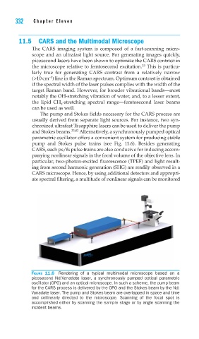
pump and Stokes pulse trains (see Fig. 11.6). Besides generating
CARS, such ps/fs pulse trains are also conducive for inducing accom-
panying nonlinear signals in the focal volume of the objective lens. In
particular, two-photon-excited fluorescence (TPEF) and light result-
ing from second harmonic generation (SHG) are readily observed in a
CARS microscope. Hence, by using additional detectors and appropri-
ate spectral filtering, a multitude of nonlinear signals can be monitored
FIGURE 11.6 Rendering of a typical multimodal microscope based on a
picosecond Nd:Vanadate laser, a synchronously pumped optical parametric
oscillator (OPO) and an optical microscope. In such a scheme, the pump beam
for the CARS process is delivered by the OPO and the Stokes beam by the Nd:
Vanadate laser. The pump and Stokes beam are overlapped in space and time
and collinearly directed to the microscope. Scanning of the focal spot is
accomplished either by scanning the sample stage or by angle scanning the
incident beams.