
Page 47 - Vibrational Spectroscopic Imaging for Biomedical Applications
P. 47
24 Cha pte r O n e
of adjacent normal and cancer TMA core datasets. These confidence
bands also reflect the narrow widths for the offset distributions for
both classes, particularly for the normal class.
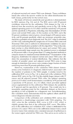
The trade off between sensitivity and specificity is demonstrated
by ROC analysis (Fig. 1.8c and d). The ROC curve with 95 percent
confidence intervals for the calibration TMA dataset in Fig. 1.8c is
derived from the sensitivity and specificity curves in Fig. 1.8b. At the
optimal operating point of a 0.3 offset cutoff the sensitivity is 93 percent
and the specificity is 94 percent, indicating clear discrimination of both
cancer and normal TMA cores. At this location on the ROC curve the
95 percent confidence interval gives a lower bound of 85 percent sensi-
tivity and 86 percent specificity, which are minimum acceptable stan-
dards for a potential cancer diagnostic tool. The AUC value is calculated
as 0.96 ± 0.02, indicating that the sample size of 31 tumor TMA cores and
34 normal TMA cores is sufficient to demonstrate confidence in cancer
53
and normal classification potential with this algorithm. Due to the min-
imal overlap in offset distributions for cancer and normal TMA cores
and the reasonably large sample size, the statistical power is calculated
as 100 percent with a z-score greater than 3.72 using standard methods to
52
compare means for standard normal distributions. Examination of the
frequency distribution for adjacent normal and cancer TMA cores vali-
dates this assumption of normal distribution. This indicates that the
number of sampled cancer and adjacent normal TMA cores is large
enough to determine that cancer cores contain a larger fraction of boxes
containing more than 50 percent epithelium pixels.
However, examination of the validation TMA dataset demon-
strates some limitations in diagnostic determination associated with
the number of patients in the TMA sample. A similar trend to the
calibration ROC curve in Fig. 1.8c is observed in the validation TMA
dataset ROC curve in Fig. 1.8d. For this slightly larger dataset with 37
cancer and 40 adjacent normal TMA cores, the optimal operating
point at a 0.5 offset cutoff demonstrates a sensitivity of 95 percent and
a specificity of 98 percent. At a 0.3 offset, which provided optimal
cancer segmentation for the calibration TMA dataset, the sensitivity
is 97 percent and the specificity is 85 percent. This would also be a
reasonable operating point for the validation dataset, as this lower
specificity is still acceptable for a diagnostic test. Notwithstanding,
this validation study demonstrates that the optimal offset cutoff for
cancer diagnosis remains uncertain.
Comparison of the calibration and validation ROC curves also
demonstrates a disadvantage associated with TMA sampling. The
AUC value for the validation ROC curve is 0.99 ± 0.01, which is
slightly greater than that of the calibration ROC curve. This differ-
ence in the calibration and validation AUC values is attributed to the
presence of a cancer TMA core in the calibration dataset from a small
invasive tumor that contains only a minimal amount of epithelium in
the tumor region selected for TMA core. This serves as an outlier in