
Page 259 - Vogel's TEXTBOOK OF QUANTITATIVE CHEMICAL ANALYSIS
P. 259
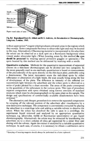
THlN-lAVER CHROMATOCRAPHV   8.6

              <
       Knob-
       Lid ---

       Plate in the-


       Fig. 86  Reproduced from D. Abbott and R. S. Andrews, An Zntroakction to Chromatography,
       Longman, London, 1965.

       with an appropriate* reagent which produces coloured areas in the regions which
       they occupy. Some compounds fluoresce in ultraviolet light and may be located
       in this way. Alternatively if fluorescing material is incorporated in the adsorbent
       the solute can  be  observed as a dark  spot on a fluorescent  background  when
       viewed  under  ultraviolet  light. (When locating zones  by  this method  the eyes
       should  be  protected by  wearing special  protective  goggles or spectacles.) The
       spots located by  this method can be delineated by marking with a needle.
       Quantitative evaluation.  Methods for the quantitative measurement of separated
       solutes on a  thin-layer  chromatogram can be  divided  into two  categories.  In
       the more generally used in-situ methods, quantitation is based on measurement
       of the photodensity of the spots directly on the thin-layer plate, preferably using
       a  densitometer.  The  latter  instrument  scans  the  individual  spots  by  either
       reflectance or absorption of a light beam, the scan usually being along the line
       of  development  of  the  plate.  The  difference in  intensity  of  the  reflected  (or
       transmitted) light between the adsorbent and the solute spots is observed  as a
       series of peaks plotted  by a chart recorder. The areas of the peaks correspond
       to the quantities of the substances in the various spots. This type of procedure
       requires comparison  with  spots obtained  using  known  amounts of  standard
       mixtures which must be chromatographed on the same plate as the sample. The
       design  and  specifications  of  commercially  available  densitometers  have  been
       re~iewed.~~
         The alternative, and cheaper, procedure is to remove the separated components
       by  scraping off  the relevant  portion  of  the  adsorbent  after  visualisation  by  a
       non-destructive technique. The component is conveniently extracted by placing
       the adsorbent in a centrifuge tube and adding a suitable solvent to dissolve the
       solute. When  the  solute  has  dissolved  the  tube  is  spun  in  a  centrifuge,  the
       supernatant  liquid  removed  and  analysed  by  an  appropriate  quantitative
       technique, e.g.  ultraviolet,  visible  or fluorescence  spectrometry  or gas-liquid
       chromatography. Alternatively  the solute may be extracted by transferring the
       adsorbent  on to a  short column of silica gel  supported  by  a  sinter filter and
       eluting with the solvent. Again the extract is analysed by a suitable quantitative
       technique. In each case, of course, it is necessary to obtain a calibration curve

       * Spraying of locating reagents should always be  performed in a fume cupboard.