
Page 134 - Advances in Biomechanics and Tissue Regeneration
P. 134
130 7. MULTISCALE NUMERICAL SIMULATION OF HEART ELECTROPHYSIOLOGY
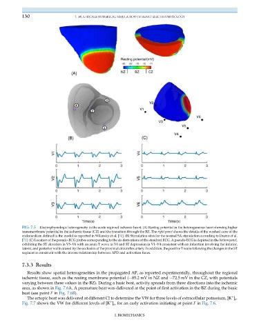
FIG. 7.5 Electrophysiologic heterogeneity in the acute regional ischemic heart. (A) Resting potential in the heterogeneous heart showing higher
transmembrane potential in the ischemic tissue (CZ) and the transition through the BZ. The right panel shows the details of the washed zone at the
endocardium defined in the model as reported in Wilensky et al. [31]. (B) Stimulation sites for the normal SA stimulation according to Durrer et al.
[71]. (C) Location of the pseudo-ECG probes corresponding to the six derivations of the standard ECG. A pseudo-ECG is depicted in the bottom panel,
exhibiting the ST elevation in V5–V6 with an acute T wave in V6 and ST depression in V1–V4 consistent with an infarction involving the inferior,
lateral, and posterior walls caused by the occlusion of the proximal circumflex artery. In addition, the positive T wave following the changes in the ST
segment is consistent with the inverse relationship between APD and activation times.
7.3.3 Results
Results show spatial heterogeneities in the propagated AP, as reported experimentally, throughout the regional
ischemic tissue, such as the resting membrane potential ( 85.2 mV in NZ and 72.5 mV in the CZ, with potentials
varying between these values in the BZ). During a basic beat, activity spreads from three directions into the ischemic
area, as shown in Fig. 7.6A. A premature beat was delivered at the point of first activation in the BZ during the basic
beat (see point F in Fig. 7.6B).
+
The ectopic beat was delivered at different CI to determine the VW for three levels of extracellular potassium, [K ] o .
+
Fig. 7.7 shows the VW for different levels of [K ] o , for an early activation initiating at point F in Fig. 7.6.
I. BIOMECHANICS