
Page 135 - Advances in Biomechanics and Tissue Regeneration
P. 135
7.3 VULNERABILITY IN REGIONALLY ISCHEMIC HUMAN HEART: EFFECT OF THE EXTRACELLULAR POTASSIUM CONCENTRATION 131
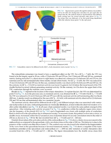
FIG. 7.6 Depolarization pattern through the ischemic tissue (delim-
ited by the white line) during basic activation (A), and right after the
ectopic beat (B). The ischemic tissue is depolarized by three different
fronts coming from the normal activation sites shown in Fig. 7.5.
The ectopic beat was delivered at the first point being depolarized
within the ischemic tissue (point F in the right panel).
+
FIG. 7.7 Vulnerability window for different levels of [K ] o . Early stimulation starts at point F in Fig. 7.6.
+
The extracellular potassium was found to have a significant effect on the VW. For a [K ] o ¼ 7 mM, the VW was
found to be the largest, equal to 20 ms, with a CI between 250 and 270 ms. For CI between 250 and 265 ms, sustained
reentry (lasting more than 3 s, about seven to eight beats) was observed, whereas for CIs between 265 and 270 ms, the
+
reentrant activity self-terminated after three reentry circuits (three beats). For [K ] o ¼ 8 mM, the VW was reduced to
15 ms (CIs between 255 and 270 ms). In this case, sustained reentry was observed for CIs between 255 and 265 ms. On
+
the contrary, for [K ] o ¼ 9.0 mM no reentrant activity was found. In general, for CIs below the lower bound of the VW,
double blocked occurred without generating reentrant activity. On the contrary, for CIs above the upper limit of the
VW, conduction through the ischemic zone occurred.
Results in Fig. 7.7 depend on the location of the ectopic stimulation. To explore this fact, the VW was determined for
+
+
point F1in Fig. 7.6. As in the previous case, no reentrant activity was obtained for [K ] o ¼ 9.0 mM. For [K ] o ¼ 7.0 mM
the VW was reduced to 10 ms (CI between 265 and 275 ms) with the reentrant activity self-terminating after two beats
+
(800 ms). A similar result was obtained for the case of [K ] o ¼ 8.0 mM, with reentrant activity observed for CIs between
290 and 310 ms that self-terminated after two reentrant circuits.
+
The reentrant activity observed for different levels of [K ] o and different ectopic sites was associated with ventric-
ular tachycardia in all cases, without degeneration in ventricular fibrillation, at least within the observed window of 3 s
after earlier stimulation (see Fig. 7.8). In all cases where sustained reentry was found, the same pattern emerged at the
epicardium. After initiating early activity (at point F in Fig. 7.6), conduction within the ischemic region slows down
(see the frequency of the pseudo-ECG in Fig. 7.8C) and a fragmented wave front with multiple areas of conduction
block is observed in the epicardium due to intramural reentry. In successive beats, this pattern evolves into a circus, or
double circus, movement within the CZ around an area of diameter about 4.0 cm and a revolution time in the order of
+
250 ms as shown in Fig. 7.8B for the last recorded beat ([K ] o ¼ 7.0 mM and CI ¼ 260 ms).
Detailed observation of the activation activity at the endocardium in Fig. 7.8A shows a faster circus movement in the
endocardium as compared to the epicardium, with a revolution time on the order of 200 ms. This behavior is due to the
presence of the washed zone that allows faster CV. The washed zone was also found to be related to the fragmentation
of the wave front at the epicardium because it favored transmural reentry. Fig. 7.9A shows a snapshot of the trans-
membrane potential at the epicardium and endocardium at t ¼ 2500 ms after initiation of earlier stimulation (last
+
recorded beat) for a [K ] o ¼ 7.0 mM and CI ¼ 260 ms. The figure demonstrates how the double circus closes first
I. BIOMECHANICS