
Page 150 - Artificial Intelligence for Computational Modeling of the Heart
P. 150
122 Chapter 4 Data-driven reduction of cardiac models
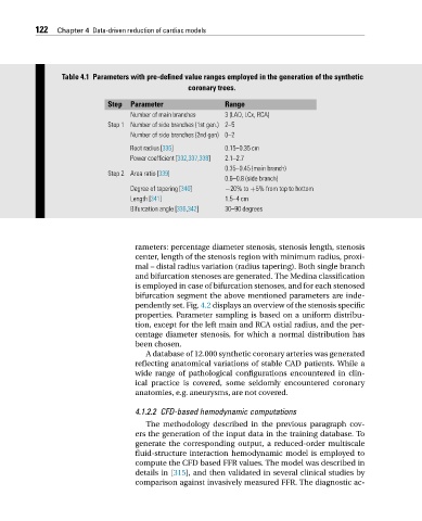
Table 4.1 Parameters with pre-defined value ranges employed in the generation of the synthetic
coronary trees.
Step Parameter Range
Number of main branches 3 (LAD, LCx, RCA)
Step 1 Number of side branches (1st gen.) 2–5
Number of side branches (2nd gen) 0–2
Root radius [336] 0.15–0.35 cm
Power coefficient [332,337,338] 2.1–2.7
0.35–0.45 (main branch)
Step 2 Area ratio [339]
0.6–0.8 (side branch)
Degree of tapering [340] −20% to +5% from top to bottom
Length [341] 1.5–4 cm
Bifurcation angle [336,342] 30–90 degrees
rameters: percentage diameter stenosis, stenosis length, stenosis
center, length of the stenosis region with minimum radius, proxi-
mal – distal radius variation (radius tapering). Both single branch
and bifurcation stenoses are generated. The Medina classification
is employed in case of bifurcation stenoses, and for each stenosed
bifurcation segment the above mentioned parameters are inde-
pendently set. Fig. 4.2 displays an overview of the stenosis specific
properties. Parameter sampling is based on a uniform distribu-
tion, except for the left main and RCA ostial radius, and the per-
centage diameter stenosis, for which a normal distribution has
been chosen.
A database of 12.000 synthetic coronary arteries was generated
reflecting anatomical variations of stable CAD patients. While a
wide range of pathological configurations encountered in clin-
ical practice is covered, some seldomly encountered coronary
anatomies, e.g. aneurysms, are not covered.
4.1.2.2 CFD-based hemodynamic computations
The methodology described in the previous paragraph cov-
ers the generation of the input data in the training database. To
generate the corresponding output, a reduced-order multiscale
fluid-structure interaction hemodynamic model is employed to
compute the CFD based FFR values. The model was described in
details in [315], and then validated in several clinical studies by
comparison against invasively measured FFR. The diagnostic ac-